

International Journal of Respiratory and Pulmonary Medicine
Iatrogenic Tracheal Rupture - The Role of a Thoracic Surgeon
Nikolaos Panagiotopoulos1*, Mohamed. A Gulamhussein1, Davide Patrini1, Rajeev Shukla1, Martin Lees2, Martin Hayward1, Shyamsunder Kolvekar1 and David Lawrence1
1Cardiothoracic Surgery Department, University College London Hospitals UCLH, UK
2Department of Cardiothoracic Anesthesia, University College London Hospitals UCLH, UK
*Corresponding author: Mr Nikolaos Panagiotopoulos, Consultant Thoracic Surgeon, Cardiothoracic Surgery Department, University College London Hospitals UCLH, 16-18 Westmoreland Street- London W1G 8PH, UK, E-mail: nikolaos.panagiotopoulos@nhs.net
Int J Respir Pulm Med, IJRPM-2-032, (Volume 2, Issue 4), Case Report; ISSN: 2378-3516
Received: August 08, 2015 | Accepted: October 26, 2015 | Published: October 28, 2015
Citation: Panagiotopoulos N, Gulamhussein MA, Patrini D, Shukla R, Lees M, et al. (2015) Iatrogenic Tracheal Rupture - The Role of a Thoracic Surgeon. Int J Respir Pulm Med 2:032. 10.23937/2378-3516/1410032
Copyright: © 2015 Panagiotopoulos N, et al. This is an open-access article distributed under the terms of the Creative Commons Attribution License, which permits unrestricted use, distribution, and reproduction in any medium, provided the original author and source are credited.
Abstract
Iatrogenic tracheal rupture following intubation is a rare, but potentially lethal complication. Usually patients present with the classical symptoms of mediastinal subcutaneous emphysema and respiratory distress. However, the timing or the onset of these symptoms is quite variable and differs between patients. Both conservative and surgical methods have been advocated in the past for the management of a diagnosed tracheal laceration. We discuss the case of a 65-year-old gentleman who was admitted electively for coronary artery bypass grafting. The patient faced difficult intubation, which led to the development of a tracheal laceration and was conservatively managed. This case emphasises on the importance of early diagnosis and prompt management being the key factors in governing optimal clinical outcome.
Keywords
Tracheal rupture, Mediastinitis, Intubation complications
Introduction
Difficulties in orotracheal intubation represent one of the major causes of iatrogenic trauma to the tracheobronchial tree. These problems usually result from repeated attempts at intubation termed as 'difficult intubations'. The resultant tracheal lacerations may also be due to underlying congenital anomalies, over inflation or rupture of the tube cuff, or in some cases, in apparently uneventful intubations [1]. The underlying laceration may result in tracheobronchial disruption presenting with life threatening tension pneumothorax, mediastinal emphysema and prolapse of esophageal wall into the trachea resulting in acute asphyxia. Late complications such as mediastinitis and sepsis further complicate the clinical picture, ultimately leading to death in these patients [2]. Once diagnosed, treatment options include conservative and surgical approaches. We discuss this case to highlight the important factors and steps to be taken when considering the challenging management of iatrogenic tracheobronchial injuries.
Case presentation
A 65- year old gentleman with triple vessel atherosclerotic coronary artery disease was admitted for an elective coronary artery bypass grafting procedure. During induction to anaesthesia intubation proved to be challenging (Grade 3) and following multiple attempts it was achieved using a bougie and a video laryngoscope. The surgery was uneventful and the patient was transferred in a stable condition and intubated to the intensive care unit. On extubation the following day, the patient developed sudden shortness of breath, accompanied by widespread subcutaneous emphysema to the neck and face. He was immediately reintubated and a suspicion of tracheobronchial injury was raised.
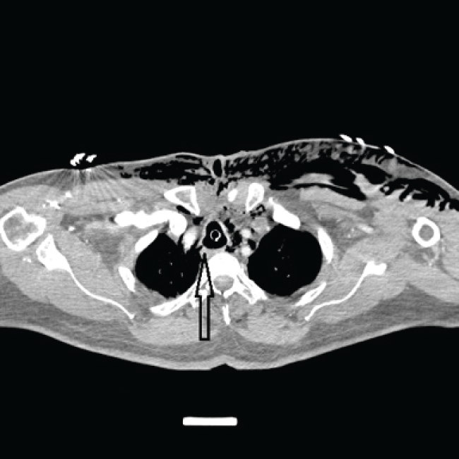
A computed tomography scan (CT scan) was then performed that confirmed the severe and widespread surgical emphysema involving the chest wall, extending around the face, back of the neck and the deep fascial planes of the neck. This was further supported by severe pneumomediastinum with air leaking into the retroperitoneum. No pneumothorax was noted. A small tracheal perforation was identified in the membranous part of the right tracheal wall 6 cm above the level of carina (Figure1) that was confirmed also by bronchoscopy. A decision was made for conservative management by keeping the patient intubated and ventilating more distally to the perforation thus bypassing the injury and hence allowing time to monitor progress of the patient's hemodynamic status.
.
Figure 1: CT neck and chest confirming the extensive surgical emphysema and showing the tracheal rupture (arrow).
View Figure 1
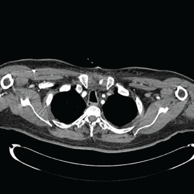
Daily chest radiographs were obtained showing progressive improvement of the surgical emphysema. Intravenous broad-spectrum antibiotics were commenced to prevent underlying mediastinitis or sepsis. The patient was weaned from the ventilator and was extubated on day 5 postoperatively. Subsequent CT showed near complete resolution of the previous widespread emphysema and no evidence of tracheal laceration (Figure 2). He had an uneventful recovery and was discharged home on day 8 in excellent clinical condition.
.
Figure 2: CT chest showing complete heal of the tracheal laceration and resolved surgical emphysema.
View Figure 2
Discussion
Tracheobronchial injury following endotracheal intubation is an infrequent, but potentially lethal entity presenting with an incidence of less than 1% [3]. Factors such as short stature, female gender, underlying medical conditions such as COPD and prolonged steroid usage remain consistent in many reports of similar cases. On the contrary, issues concerning technique of intubation such as multiple attempts, inexperienced operators, inappropriate use of stylets or cuff over inflation, malpositioning of the tube and lastly patient abrupt movement or excessive coughing comprise the remaining issues associated with the underlying mode of injury [4,5].
Of note however is the site of tracheal injury, which in most cases is found to be above or at the level of the carina and mostly involving the membranous portion of the trachea. Bronchoscopy is the mandatory investigation to establish the diagnosis and to identify the location and choose the appropriate treatment and approach. Nonsurgical therapy must be considered in small (< 2 cm) uncomplicated tears in stable patients as healing can be achieved in a few days with minimal risks and discomfort for the patient [6,7]. Surgery - if necessary-should be performed promptly to guarantee success, and to avoid one of the most feared complications of this injury, mediastinitis. Various surgical approaches have been identified for large lacerations (> 3 cm) ranging from a right thoracotomy approach to a transcervical approach depending on the site or location of the laceration and the room for successful sealing of the perforation considering the number of vital structures involved in the operating field [5]. The thoracotomy approach is usually incorporated while dealing with tracheal lacerations extending to the membranous part of the main bronchi. The cervical approach is used for post intubation lesions limited to the trachea. In some cases, the lesion is discovered during the surgical procedure where a primary repair is usually possible. Alternatively, for certain intermediate lesions, a tracheostomy may be employed as a means of reducing airway pressure and decreasing the air leak. However the conventional nasotracheal intubation with the cuff inflated distal to the tear is equally effective in preventing further injury to the already damaged tracheal wall [1,7].
A female predominance can be explained by the fact that females tend to be shorter and improperly large tubes tend to be selected for them. Furthermore, their tracheal diameters are noted to be smaller compared to men and they may be more prone to cuff over inflation as a result. Underlying comorbidities, skeletal anomalies and the use of vasodilator agents are all factors that need immense consideration [8,9].
Conclusion
The management options of post intubation tracheal rupture remain individualized. Early surgical repair is the preferred treatment modality in selected cases. Conservative treatment options may be a viable choice in patients with small-uncomplicated tears in the absence of gross air leaks or in patients with significant comorbidities. Even in the case of a slight suspicion, immediate investigation by chest radiography and bronchoscopy should be employed to prevent the fatal aftermath involved with this form of iatrogenic trauma [2,3].
Disclosure
All the authors have declared no competing interest.
References
-
Orta DA, Cousar JE 3rd, Yergin BM, Olsen GN (1979) Tracheal laceration with massive subcutaneous emphysema: a rare complication of endotracheal intubation. Thorax 34: 665-669.
-
Kaloud H, Smolle-Juettner FM, Prause G, List WF (1997) Iatrogenic ruptures of the tracheobronchial tree. Chest 112: 774-778.
-
Borasio P, Ardissone F, Chiampo G (1997) Post-intubation tracheal rupture. A report on ten cases. Eur J Cardiothorac Surg 12: 98-100.
-
Berry M, Van Schil P, Van Meerbeeck J, Vanmaele R, Eyskens E (1997) Surgical treatment of iatrogenic tracheal lacerations. Acta Chir Belg 97: 308-310.
-
Angelillo-Mackinlay T (1995) Transcervical repair of distal membranous tracheal laceration. Ann ThoracSurg 59: 531-532.
-
Ross HM, Grant FJ, Wilson RS, Burt ME (1997) Nonoperative management of tracheal laceration during endotracheal intubation. Ann Thorac Surg 63: 240-242.
-
Roxburgh JC (1987) Rupture of the tracheobronchial tree. Thorax 42: 681-688.
-
Jougon J, Ballester M, Choukroun E, Dubrez J, Reboul G, et al. (2000) Conservative treatment for postintubation tracheobronchial rupture. Ann Thorac Surg 69: 216-220.
-
Chen EH, Logman ZM, Glass PS, Bilfinger TV (2001) A case of tracheal injury after emergent endotracheal intubation: a review of the literature and causalities. Anesth Analg 93: 1270-1271.
