

Journal of Musculoskeletal Disorders and Treatment
The Effects of an Audible Low Frequency Acoustic Waveform on Osteoarthritis: A Pilot Study
Harvey W Wallmann* and William R VanWye
Department of Physical Therapy, Western Kentucky University, Kentucky, USA
*Corresponding author:
Harvey W Wallmann, PT, DSc, SCS, ATC, CSCS, Professor, Head, Department of Physical Therapy, The Medical Center - WKU, Western Kentucky University, Health Sciences Complex, 700 First Avenue, Bowling Green, KY 42101, USA, Tel: (270) 745-4070, Fax: (270) 745-3497, E-mail: harvey.wallmann@wku.edu
J Musculoskelet Disord Treat, JMDT-2-021, (Volume 2, Issue 3), Research Article
Received: June 20, 2016; Accepted: September 12, 2016; Published: September 15, 2016
Citation: Wallmann HW, VanWye WR (2016) The Effects of an Audible Low Frequency Acoustic Waveform on Osteoarthritis: A Pilot Study. J Musculoskelet Disord Treat 2:021.
Copyright: © 2016 Wallmann HW, et al. This is an open-access article distributed under the terms of the Creative Commons Attribution License, which permits unrestricted use, distribution, and reproduction in any medium, provided the original author and source are credited.
Abstract
Objective:The purpose of this pilot study was to investigate the effects of an audible low frequency acoustic waveform on pain and range of motion (ROM) for patients with osteoarthritis (OA).
Methods:Twenty one adults with OA (7 males and 14 females with a mean age of 68.1 ± 12.4) participated in the study and were recruited from local advertisements to participate in a quasi-experimental pre-test, post-test, 24 hour post-test design using a new technology called the Medsonix Therapy System. Prior to and after the intervention, ROM was measured for the wrist, knee, and hip using goniometry, and pain was assessed using a visual analog pain scale across all conditions. Six participants at a time were then seated in a circle facing a column housing a generator approximately one foot away from the column and were given the intervention for 25 minutes. Data were analyzed using one-way repeated measures ANOVA.
Results:Patients had less pain immediately (p < 0.001) and at 24 hours (p < 0.01). ROM significantly improved in right and left hip flexion (p < 0.01), left wrist flexion (p < 0.01), and left knee flexion (p < 0.05) pre to post. Significant improvements were noted in hip flexion (left, p < 0.001; right, p < 0.01) and wrist flexion (left, p < 0.05; right, p < 0.01) after 24 hours, but not in knee flexion or wrist extension ROM.
Conclusion:The results of this pilot study suggest that use of the Medsonix Therapy System as an alternative form of therapy appears to improve ROM in various joints while decreasing pain in individuals diagnosed with OA utilizing an audible low frequency acoustic waveform.
Keywords
Alternate therapy, Osteoarthritis, Audible sound, Low frequency waveform, Pain
Introduction
Osteoarthritis (OA), referred to as degenerative joint disease, [1,2] affects over 30 million U.S. adults, [3,4] accounting for billions of dollars spent on joint replacements [5] and lost work. Modifiable risk factors for developing OA include excessive body mass, joint injury, muscle weakness, and occupation as well as non-modifiable factors such as female gender, increasing age, and genetic predisposition [6-11].
Methods of treating pain in OA include surgical, pharmacological, and non-pharmacological interventions. Recommendations for pharmacological management of OA include oral nonsteroidal anti-inflammatory drugs, acetaminophen, and tramadol. Although intra-articular corticosteroid injections are recommended for the knee and hip, they are not for the hand [12]. Complementary and alternative treatments (CAM) such as herbal remedies, supplements, and diet therapies have also been used in attempts to treat OA. However, even commonly used CAM such as chondroitin sulfate and glucosamine, have not been substantiated by medical research for use to manage OA and have sometimes proven to be very costly to the patient [12,13]. Strong recommendations for non-pharmacological management of OA include weight loss, aerobic, resistance, and aquatic exercise.
Management of OA via invasive procedures or pharmacological treatment are not without side effects. Therefore, investigation of non-pharmacological and CAM treatments, which often have less side-effects would appear prudent. One new technology that may provide pain relief for patients with OA is the use of audible sound technology. This technology, the Medsonix Therapy System, is registered with the FDA as a Class 1 Medical Device (low risk) and has been granted three distinct U.S. patents for use as a low frequency acoustic methodology for treating pain [14].
Recent anecdotal reports have been received demonstrating that audible sound in a particular range below 1000 Hz has had astounding effects on reducing the pain associated with OA. For example, a previous investigation of this technology found the effects of a low frequency acoustic waveform (audible sound) on peripheral vascular disease (PVD) and found a significant increase in the right ankle brachial index (RABI) as well as increases in blood flow in several of the arteries assessed [14]. However, there are currently no studies that have addressed the use of audible sound as a treatment for OA. Due to the paucity of research, there is a need to investigate the relationship between audible sound in this frequency range and its effect on disease states. Information derived from empirically measuring outcomes such as pain and ROM might help to further elucidate the mechanisms involved with low frequency audible sound. Using the Medsonix Therapy System, the purpose of this study was to examine the effects of an audible low frequency acoustic waveform on OA. It is hypothesized that the intervention using this technology will result in increased ROM and decreased pain in a population of individuals suffering from OA.
Methods
Participants
This quasi-experimental research study utilized a one group repeated measures pretest-posttest design. The authors have no financial or other interest in the product or distributor of the product. Prior to engaging in the study, the university’s biomedical institutional review board approved the study and informed oral and written consents were obtained from eligible participants. Seven males and 14 females with a mean age of 68.1 ± 12.4 years participated in the study and were recruited through local newspaper advertisements. Interested participants attended a 30-minute information session prior to the study to determine their eligibility, which included a previously diagnosed OA condition, English speaking, no previous sonic therapy treatments, no implanted devices, and being non-pregnant. It was not determined whether participants had any previous OA treatment or OA-related surgery. Participants were informed not to change their normal daily routines. Researchers emphasized the importance of continuing usual patterns of activity and exercise, diet, hydration, rest, and medications prior to the study.
Equipment
The treatment was performed using a new technology called the Medsonix Therapy System (Figure 1), which consists of a control unit and the Medsonix transducer. The Medsonix transducer is controlled by the Medsonix Therapy System control unit which operates the transducer at a specific frequency, amplitude, and time.
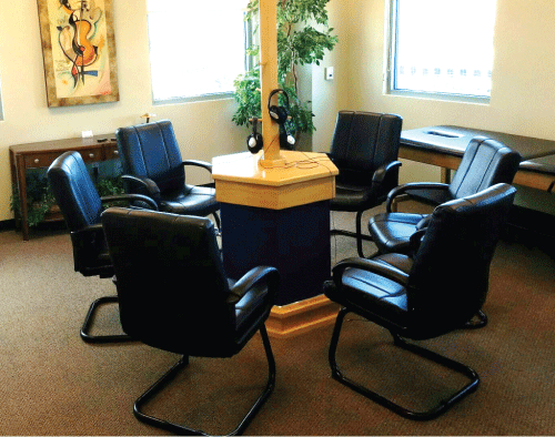
A key facet of this technology is the efficiency in which electrical energy is converted to mechanical movement. The Medsonix transducer is capable of operating at high efficiency, while resonating at a specified low frequency. Additionally, it resonates with an omnidirectional beam pattern. The Medsonix transducer emits a moderately loud sound, exposing participants to approximately 80 decibels of sound (without the headphones on), which is within OSHA standards. Use of the headphones effectively reduces the decibels of sound to which the participant is exposed. The Medsonix Therapy System operates within the range of 400-800 Hz with an optimum frequency of approximately 600 Hz.
Visual analog pain scale
A visual analog pain scale (VAS) was utilized to determine OA pain perception before the intervention, after the intervention, and 24-hours later. The scale was a standard 100 mm line ranging from no OA pain to worst OA pain ever. Participants were asked to mark through the line in accordance with how much pain was felt at that time. Researchers were able to accurately measure the amount of pain indicated with the use of a millimeter ruler [15]. Researchers have shown that when examiners use the same pain scales when assessing and reassessing patient’s perceived pain, valid and reliable results have been achieved [16-18]. A minimal clinically important difference (MCID) for patients with knee and hip OA has been established for the VAS, yet it varies depending on baseline pain scores. Baseline scores of 30-49 mm would require 7 mm of change and baseline scores of 50-65 mm would require 24 mm of change for hip and knee OA [19].
Goniometry
Many organizations including the American Medical Association and the American Academy of Orthopaedic Surgeons and researchers have adopted the 0 to 180 degree goniometric notation system as an objective measurement of joint positions [20,21]. Intra-rater reliability of active goniometric measurements of the hip, knee, and wrist has been shown to be good to excellent [22,23]. Accuracy of goniometric measurements can be improved if taken by a single clinician [24]. Based on this principle, only one clinician performed pre-treatment and post-treatment passive ROM measurements using the 0 to 180 degree goniometric notation system at the wrist and metacarpal phalangeal joints bilaterally. To avoid any bias of the clinician performing the measurements, a different individual recorded the values of the measurements as stated by the clinician. Goniometric measurements were performed pre-treatment, post-treatment, and 24 hours post-treatment.
Procedure
On the day of the study prior to the intervention, participants completed a brief background questionnaire along with a visual analog pain scale. Body areas measured were bilateral wrist extension, wrist flexion, knee flexion, and hip flexion. The data were separated into right and left goniometric outcome measures as well as pre, post, and 24-hour measures of pain. Following these pre-measurements, participants were escorted to a room for the treatment. Up to six participants were treated at one time and were seated facing the Medsonix transducer in a circular fashion approximately one foot away from the generator column. Participants were asked to wear headphones through which relaxation music could be controlled by individual volume dials. The treatment consisted of sitting in a chair for 25 minutes during which time the Medsonix transducer was turned on. Post measurements were taken for the same variables following the treatment and follow-up measurements were taken at 24 hours.
Data analysis
Visual analog and goniometric data were analyzed using one-way repeated measures ANOVA (SPSS, version 23.0). When the omnibus F-ratio was significant, planned comparisons were conducted with the alpha level set at 0.05 using the main effects comparison option available via SPSS. The planned comparisons of interest were the comparisons of ROM and pain between measurement times.
Results
Comparisons of pre, post, and 24-hour data for all 21 participants for each of the body areas examined are summarized in (Tables 1, Table 2 and Table3).
![]()
Table 1: Outcome measures for right wrist, knee, and hip across time.
View Table 1
![]()
Table 2: Outcome measures for left wrist, knee, and hip across time.
View Table 2
The means of all participants demonstrated increased ROM changes over a 24-hour period for all of the areas examined. Right hip flexion measurements differed, F2,40 = 11.426, P < 0.0005 with increased measurements noted pre to post (p = 0.004) and pre to 24-hour (p = 0.001). Left hip flexion measurements changed as well, F2,40 = 22.636, P < 0.0005 with increased measurements noted pre to post (p = 0.000) as well as pre to 24 hour (p = 0.000). Significant differences were also observed for left wrist flexion, F2,40 = 5.332, P = 0.009 with increased measurements demonstrated pre to post (p = 0.002) and pre to 24-hour (p = 0.019). Right wrist flexion measurements, F2,40 = 6.424, P = 0.004 only increased pre to 24-hour (p = 0.004). Changes were also observed for knee flexion, F2,40 = 4.347, P = 0.02 with significant differences noted pre to post (p = 0.010). Additionally, participants demonstrated significant decreases in pain, F2,40 = 13.911, P < 0.0005 with differences noted pre to post (p = 0.000) as well as pre to 24-hour (p = 0.007). Observed power ranged from 72.1-100%.
![]()
Table 3: Outcome measures for pain across time.
View Table 3
Discussion
The present study was designed to evaluate the effects of an audible low frequency acoustic waveform on OA and to determine whether or not it could be a useful treatment for sufferers of this disease. This was conducted in order to provide a necessary background for further research with this device for OA and other disease states. All of the participants in this study showed improvement over a 24-hour period. It was demonstrated that exposure to the audible low frequency acoustic waveforms at the designated frequency significantly increased the ROM in several of the body areas examined immediately post treatment as well as 24 hours post treatment. Additionally, the intervention significantly decreased pain immediately post treatment and 24-hours post treatment. Given the MCID for baseline changes of 50 to 65 mm (5 to 6 cm), the necessary decrease in pain would need to be 24 mm (2.4 cm). Since the immediate post treatment pain decreased 2.53 cm, this represents a true decrease in pain. The pre to 24 hour pain measurement, although statistically significant, may not be clinically significant if the pain stemmed mainly from the hip and knee (only a 1.31 cm change was observed); but since a global pain rating was taken (pain was not assessed for specific joints), this cannot be definitively determined. No untoward side effects were noted. We are unable to corroborate the results obtained with other studies because of the novelty of this intervention. We are also unable to unequivocally explain why the intervention led only to improvements in flexion and not left and right wrist extension at this time except that perhaps the observed power was considerably less for these two ROMs.
Studies involving infrasound (i.e. under 16 Hz) have yielded diverging results and have mostly explored the effects of on the auditory and non-auditory systems [25-29]. Although therapeutic US has achieved recognition as a suitable method to treat a wide variety of musculoskeletal conditions, those conducted on individuals suffering with OA have been unable to distinguish its effects from sham therapy [30]. The characteristics of audible low frequency acoustic waveforms involved in physiological reactions aside from the auditory system are largely unknown. With the exception of the PVD study, no studies to date have examined the effects of full body exposure to low frequency audible sound in this range for any disease state [14].
Sound waves are a form of vibration and are divided into three groups: infrasound, audible sound, and ultrasound. Infrasound ranges from 1-16 Hz (inaudible), audible sound ranges from 16-20,000 Hz, and therapeutic ultrasound (inaudible) is any sound wave with a frequency above 20,000 Hz [31]. Ultrasound has been shown to have thermal and non-thermal effects on the body. Although many studies involving the use of ultrasound exist, there are relatively few studies concerned with the physiological effects on humans of infrasound exposure [32]. Audible sound is, in contrast to all other environmental factors, continuously present in the external environment. Audible sound has been shown to have physiological effects on the body and its metabolic processes by activating subcortical neural systems. By activating these systems, the cardiovascular, metabolic, endocrine, reproductive, and neurological functions of the body may be altered [33].
A study performed by Jensen and Rasmussen [34] exposed mice to a sound at 800 Hz and an intensity between 120-123 decibels. Exposing the mice to the sound for 3 hours daily for 30 days was shown to impair interferon production and limit the inflammatory response. They found that the stress caused by the sound induced hyperactivity of the pituitary-adrenocortical axis, which they believe to be the cause of the inhibition of the inflammatory response. Henkin and Knigge [35] showed similar results, exposing rats to 220 Hz at 130 decibels for up to 48 hours. Their research showed that this intense sound doubled the output of corticosterone in 30 minutes and tripled the output in 60 minutes. Billewicz-Stankiewicz and Krepinska-Urban [36] also reported an inhibition of the inflammatory response after exposing rats to 2 hours of 86 dB sound (sound and vibration).
In 1981, Borg investigated the physiological and pathogenic effects of sound [27]. His study involved exposing rats to environmental noise for 10 hours per day for the life span of the rat at levels of 85 and 105 decibels. Findings showed that there were no significant changes in blood pressure, body weight, water consumption, life span, or disease panorama. The only potential risks determined were hearing loss and lesions to the sensory cells of the inner ear. These were related to exposure level, duration of exposure, and the strain of the animal. Borg concluded that exposure to “pseudo-constant neutral” sound at this intensity poses no threat to the health of humans except for a potential loss of hearing sensitivity in those studies that use excessively high decibel levels for prolonged periods of time.
Our study has several limitations. First, we realize this study is subject to recruitment bias. All of the participants in this study responded to a solicitation to participate in a trial of audible low frequency acoustic waveforms treatment for OA. Second, although the participants stated they all suffered with OA and that they had been previously diagnosed with the condition, it is possible that some participants may have had other conditions that mimicked OA and were unintentionally used in our study. Third, in order to prove a treatment efficacious, the prescription of treatment for OA should follow general guidelines of medical therapy. Due to the novelty of this treatment, indications, dosage regimens, and other therapeutic effects have yet to be determined. Fourth, the small number of participants and the lack of a control group limit the current study. Fifth, since we emphasized that the participants continue with their usual patterns of activity to include taking medications if needed and did not identify whether participants had any previous OA treatment or OA-related surgery, we are unable to determine any effects this may have had. Lastly, we also realize that the music may have had a relaxing effect on the participants, which may have served to decrease pain immediately post-treatment. However, this does not explain the continued decrease in pain 24 hours later. Additionally, changes in goniometric measurement may have simply been due to measurement error.
Despite the obvious limitations, we believe that this study adds pertinent information regarding the use of an alternative therapy for patients experiencing OA. No attempt was made to elucidate a mechanism of action at this time. Future research on other disorders is currently being conducted using this device. Based on the promising results of this study and the previous study on PVD, research is being planned to use double blind, randomized control designs in an effort to distinguish the efficacy of this intervention from that of a placebo effect on not only PVD and OA, but also rheumatoid arthritis.
Conclusion
The results of this pilot study suggest that use of the Medsonix Therapy System as an alternative form of therapy appears to improve ROM in various joints, while decreasing pain in individuals diagnosed with OA utilizing an audible low frequency acoustic waveform. These results are sufficiently positive to warrant more definitive research concerning empirical data as well as attempting to establish a mechanism of action using this device.
Acknowledgement
The authors wish to thank Alphonse Cassone for his technical assistance and the use of his patented technology.
References
-
Lawrence RC, Felson DT, Helmick CG, Arnold LM, Choi H, et al. (2008) Estimates of the prevalence of arthritis and other rheumatic conditions in the United States. Part II. Arthritis Rheum 58: 26-35.
-
Cisternas MG, Murphy L, Sacks JJ, Solomon DH, Pasta DJ, et al. (2016) Alternative Methods for Defining Osteoarthritis and the Impact on Estimating Prevalence in a US Population-Based Survey. Arthritis Care Res (Hoboken) 68: 574-580.
-
Buckwalter JA, Saltzman C, Brown T (2004) The impact of osteoarthritis: implications for research. Clin Orthop Relat Res S6-15.
-
Murray CJ, Atkinson C, Bhalla K, Birbeck G, Burstein R et al. (2013) The state of US health, 1990-2010: burden of diseases, injuries, and risk factors. JAMA 310: 591-608.
-
Murphy L, Helmick CG (2012) The impact of osteoarthritis in the United States: a population-health perspective: A population-based review of the fourth most common cause of hospitalization in U.S. adults. Orthop Nurs 31: 85-91.
-
Blagojevic M, Jinks C, Jeffery A, Jordan KP (2010) Risk factors for onset of osteoarthritis of the knee in older adults: a systematic review and meta-analysis. Osteoarthritis Cartilage 18: 24-33.
-
Cooper C, Snow S, McAlindon TE, Kellingray S, Stuart B, et al. (2000) Risk factors for the incidence and progression of radiographic knee osteoarthritis. Arthritis Rheum 43: 995-1000.
-
Felson DT (2004) Risk factors for osteoarthritis: understanding joint vulnerability. Clin Orthop Relat Res S16-21.
-
Felson DT, Zhang Y (1998) An update on the epidemiology of knee and hip osteoarthritis with a view to prevention. Arthritis Rheum 41: 1343-1355.
-
Jordan JM, Helmick CG, Renner JB, Luta G, Dragomir AD, et al. (2007) Prevalence of knee symptoms and radiographic and symptomatic knee osteoarthritis in African Americans and Caucasians: the Johnston County Osteoarthritis Project. J Rheumatol 34: 172-180.
-
Rossignol M, Leclerc A, Allaert FA, Rozenberg S, Valat J, et al. (2005) Primary osteoarthritis of hip, knee, and hand in relation to occupational exposure. Occup Environ Med 62: 772-777.
-
Hochberg MC, Altman RD, April KT, Benkhalti M, Guyatt G, et al. (2012) American College of Rheumatology 2012 recommendations for the use of nonpharmacologic and pharmacologic therapies in osteoarthritis of the hand, hip, and knee. Arthritis Care Res (Hoboken) 64: 465-474.
-
Zochling J, March L, Lapsley H, Cross M, Tribe K, et al. (2004) Use of complementary medicines for osteoarthritis--a prospective study. Ann Rheum Dis 63: 549-554.
-
Candela LL, Wallmann HW, Witt CS (2002) The effects of a low frequency acoustic waveform on peripheral vascular disease: a pilot study. Complementary Therapies in Medicine 10: 170-175.
-
Hawker GA, Mian S, Kendzerska T, French M (2011) Measures of adult pain: Visual Analog Scale for Pain (VAS Pain), Numeric Rating Scale for Pain (NRS Pain), McGill Pain Questionnaire (MPQ), Short-Form McGill Pain Questionnaire (SF-MPQ), Chronic Pain Grade Scale (CPGS), Short Form-36 Bodily Pain Scale (SF-36 BPS), and Measure of Intermittent and Constant Osteoarthritis Pain (ICOAP). Arthritis Care Res (Hoboken) 63: S240-252.
-
Carlsson AM (1983) Assessment of chronic pain. I. Aspects of the reliability and validity of the visual analogue scale. Pain 16: 87-101.
-
Langley GB, Sheppeard H (1985) The visual analogue scale: its use in pain measurement. Rheumatol Int 5: 145-148.
-
Scott J, Huskisson EC (1979) Vertical or horizontal visual analogue scales. Ann Rheum Dis 38: 560.
-
Tubach F, Ravaud P, Baron G, Logeart I, Bellamy N, et al. (2005) Evaluation of clinically relevant changes in patient reported outcomes in knee and hip osteoarthritis: the minimal clinically important improvement. Ann Rheum Dis 64: 29-33.
-
American Medical Association (1990) Guides to the evaluation of permanent impairment. (3rd edn), Milwaukee: AMA.
-
Riddle DL, Rothstein JM, Lamb RL (1987) Goniometric reliability in a clinical setting. Shoulder measurements. Phys Ther 67: 668-673.
-
Clapper MP, Wolf SL (1988) Comparison of the reliability of the Orthoranger and the standard goniometer for assessing active lower extremity range of motion. Phys Ther 68: 214-218.
-
Horger MM (1990) The reliability of goniometric measurements of active and passive wrist motions. Am J Occup Ther 44: 342-348.
-
Goodwin J, Clark C, Deakes J, Burdon D, Lawrence C (1992) Clinical methods of goniometry: a comparative study. Disabil Rehabil 14: 10-15.
-
Borg E (1979) Physiological aspects of the effects of sound on man and animals. Acta Otolaryngol Suppl 360: 80-85.
-
Borg E (1981) Noise, hearing and hypertension. Scandinavian Audiology 10: 125-126.
-
Borg E (1981) Physiological and pathogenic effects of sound. Acta Otolaryngol Suppl 381: 1-68.
-
Hammelburg E (1974) Biological effects of sound waves. Prog Biometeorol 1: 409-412.
-
Slarve RN, Johnson DL (1975) Human whole-body exposure to infrasound. Aviat Space Environ Med 46: 428-431.
-
Falconer J, Hayes KW, Chang RW (1992) Effect of ultrasound on mobility in osteoarthritis of the knee. A randomized clinical trial. Arthritis Care Res 5: 29-35.
-
Zagzebski JA (1996) Essentials of Ultrasound Physics. St. Louis: Mosby.
-
Westin JB (1975) Infrasound: a short review of effects on man. Aviat Space Environ Med 46: 1135-1140.
-
Welch BL (1970) Environmental noise, "adaptation" and pathological change. In: Welch BL & Welch AS, ed. Physiological effects of noise. New York: Plenum Press 5,6.
-
Jensen MM, Rasmussen Jr AF (1970) Audiogenic stress and susceptibility to infection. In: Welch B L & Welch AS, Physiological effects of noise. Plenum Press, New York, 7-19.
-
Henkin RI, Knigge KM (1963) Effect of sound on the hypothalamic-pituitary-adrenal axis. Am J Physiol 204: 701-704.
-
Billewicz-Stankiewicz J, Krepinska-Urban A (1974) The effect of vibration and noise on development of inflammatory reaction in rats. Acta Physiol Pol 25: 235-240.
