

Journal of Rheumatic Diseases and Treatment
Anti-inflammatory Effect of Antioxidant Pequi (Caryocar Brasiliense) Oil Capsules and Antioxidant Effect of Vitamin D and Physical Activity on Systemic Lupus Erythematosus Patients
Thaís Muniz Montalvão1,2, Ana Luisa Miranda-Vilela2,3*, Cesar Koppe Grisolia2 and Leopoldo Luiz Santos-Neto1,4
1University Hospital of Brasília, University of Brasília, Brasília, Brazil
2Department of Genetics and Morphology, Institute of Biological Sciences, University of Brasília, Brasilia, Brazil
3Department of Medicine, Faciplac, Campus Gama/DF, Brazil
4Department of Medicine, University of Brasilia, Brasília, Brazil
*Corresponding author: Ana Luisa Miranda-Vilela, Department of Genetics and Morphology, Institute of Biological Sciences, University of Brasilia, Brasilia/DF, Brazil, Tel: +55 (61) 3107-3085, Fax: +55 (61) 3107-2923, E-mail:
mirandavilela@unb.br, analuisamv@uol.com.br
J Rheum Dis Treat, JRDT-2-029, (Volume 2, Issue 1), Case Series; ISSN: 2469-5726
Received: November 26, 2015 | Accepted: January 07, 2016 | Published: January 09, 2016
Citation: Montalvão TM, Miranda-Vilela AL, Grisolia CK, Santos-Neto LL (2016) Anti-inflammatory Effect of Antioxidant Pequi (Caryocar Brasiliense) Oil Capsules and Antioxidant
Effect of Vitamin D and Physical Activity on Systemic Lupus Erythematosus Patients. J Rheum Dis Treat 2:029. 10.23937/2469-5726/1510029
Copyright: © 2016 Montalvão TM, et al. This is an open-access article distributed under the terms of the Creative Commons Attribution License, which permits unrestricted use, distribution, and reproduction in any medium, provided the original author and source are credited.
Abstract
Objectives: Verifying in SLE patients whether antioxidant supplementation with pequi oil capsules could reduce oxidative DNA damage and inflammation in SLE patients.
Patients and Methods: A controlled randomized, crossover, double-blind study was conducted with 38 SLE patients aged 18 to 60, with SLE Disease Activity Index (SLEDAI) below 10, chosen from 73 lupus patients consecutively seen in the University Hospital of Brasília (HUB) Rheumatology Clinic. They were randomized into two groups: one group of 22 patients received one placebo capsule per day for 60 consecutive days, and the other 16 patients received one 400 mg pequi oil in capsule per day over the same period. There was a 60-day wash-out, and then the patients switched groups: those who had received pequi oil received placebo and vice versa, also for 60 consecutive days. We collected blood, urine, anthropometric and social data from patients before and after each period, totaling four collections.
Results: Twenty-nine women finished this clinical trial. The mean age was 33.66 years, the mean BMI was 24.1 kg/m2 and the mean disease duration was 7.8 years. The irreversible damage index of the disease and the SLEDAI had no influence on DNA damage index (DDI). A correlation was found between DDI and HDL (r = −0.414, p = 0.029). A significant difference was detected in the average rate of DDI between physical activity practitioners and non-practitioners (p = 0.045) and between users and non-users of vitamin D (p = 0.006). Moreover, high-sensitivity C-reactive protein (hs-CRP) fell significantly after treatment with pequi oil (p = 0.0161).
Conclusions: Although pequi oil did not significantly reduce the DNA damage index, it effectively reduced hs-CRP levels in these patients, indicating reduced inflammation with the use of antioxidant supplementation. Furthermore, the practice of physical activity and the use of vitamin D in patients with SLE could have an independent antioxidant effect.
Keywords
Systemic Lupus Erythematosus, Antioxidant, Oxidative stress, Inflammatory marker, High-sensitivity C-reactive protein (hs-CRP), DNA damage, Comet assay, Pequi oil. Crossover
Introduction
Systemic lupus erythematosus (SLE) is a chronic inflammatory, complex, multisystem autoimmune disease of multifactorial origin. It is characterized by a large set of antibodies, especially antinuclear antibodies. Its incidence rate varies from 1 to 10 per 100,000 persons/year and its prevalence rate is 20-70 per 100,000. It is a more frequent rare disease in women between 15 and 45 years old [1-4]. The cause of SLE is still unknown; however, there is a loss of the regulatory mechanisms which maintain self-tolerance to nuclear antigens, resulting in the deposition of immune complexes in tissues [2,5]. SLE expression is also influenced by non-genetic factors, such as environment, ultra-violet light, smoking and sex hormones, since the frequency of this disease is 10 times higher in women during the reproductive years than in men. Thus, the interactions of genetic, hormonal and environmental factors act together to activate helper T cells and B cells, causing the production of various autoantibodies [1,2,6].
Oxidative stress is also important in the pathophysiology of autoimmune diseases because reactive oxygen species (ROS) and reactive nitrogen species attack macromolecules such as carbohydrates proteins, lipids and DNA, causing cell damage [7]. The increase in production of these species may favor the development of SLE, since oxidative stress causes immune modulation [8]. This imbalance can contribute to impairing immune cells, producing autoantigens and autoantibody reactivity. Moreover, proteins modified by oxidation are responsible for additional disorders in autoimmune diseases, since they represent potential targets for the immune system by breaking B cell tolerance [9]. Chronic inflammation is also associated with increased oxidative stress [10] and patients with SLE are in a state of chronic inflammation in the course of the disease. Furthermore, oxidative stress also contributes to chronic inflammation of the tissue [8]. There are several secondary consequences of oxidative stress in patients with SLE, such as its association with the atherogenic lipid profile [11]. This state plays a central role in dyslipidemia and atherosclerosis [8] and it is an important mediator of hypertension-mediated autoimmune mechanisms [12].
The comet assay is a rapid, simple, visual and sensitive technique widely used to measure and analyze DNA damage [13,14]. It serves to detect both simple and double-strand breaks [15,16] in mammalian cells at low cost [17] including in in vivo systems [18]. We showed in a previous study that DNA damage index (DDI) by comet assay rose in SLE patients when compared with healthy individuals, which reflects the oxidant/antioxidant imbalance in these patients, independently of disease activity. These findings support an association between oxidative stress and SLE [19].
Dietary factors are important regulators of immune function [20], and a diet with moderate protein and energy content, rich in antioxidants like vitamin E, vitamin A, selenium, calcium, mono and polyunsaturated fatty acids/omega 3 can promote a protective effect against tissue damage and inflammatory activity, besides helping treat comorbidities and adverse medicine reactions. Thus, some recommendations may offer a better quality of life to SLE patients [21,22], and using antioxidants may protect against the development of SLE by combating oxidative stress [8]. The species Caryocar brasiliense Camb., known as pequi, is a typical tree of the Brazilian Cerrado biome. All its parts are widely used, especially the fruit [23]. Besides lipids (including fatty acids, such as linolenic), protein, fibers, carbohydrates, the pequi contains, antioxidants such as carotenoids, including β-carotene, lycopene, ζ-carotene, criptoflavona, anteroxantina, zeaxanthin, mutatoxantina, violoxantina, lutein and neoxanthin, besides vitamin E, and phenolic compounds (flavonoids and tannins) [24,25]. A study of healthy street racers of both genders who received 14 days’ supplementation of 400 mg of pequi oil, extracted with chloroform, showed that there was a significant reduction in DNA damage measured by comet assay [26] and significant reduction of total cholesterol (TC) and low density lipoprotein (LDL) in the group aged over 45, mostly in men. The authors suggested that the oil works in reducing the inflammation caused by exercise, measured by C-reactive protein and thiobarbituric acid reactive substances (TBARS) [27].
As (1) patients with SLE have an imbalance in the oxidant/anti-oxidant system, promoting the formation of ROS [19]; (2) the antioxidant status of the plasma is directly related to diet and is defined by antioxidants such as carotenoids (the most powerful suppressors of biological singlet oxygen), flavonoids and vitamin E [20-22]; and (3) pequi contains such substances [24,25]; we aimed to verify in SLE patients whether antioxidant supplementation with pequi pulp oil capsules could reduce oxidative damage to DNA (evaluated by comet assay) and inflammation (evaluated by high-sensitivity C-reactive protein - hs-CRP). As there are secondary consequences of oxidative stress in patients with SLE, such as its association with atherogenic lipid profile [11], we also analyzed total cholesterol, low density lipoprotein, hemogram and other biochemical markers.
Patients and Methods
Patients and study design
Initially, 73 patients attended consecutively in the University Hospital of Brasília (HUB) Rheumatology Clinic from June to October 2011 were recruited. Of these, 38 individuals were selected within the inclusion criteria: diagnosis of SLE, according to the classification of the American College of Rheumatology (ACR) revised criteria for the classification of SLE [28] of both genders, between 18 and 60 years. Exclusion criteria were: patients with renal impairment (creatinine ≥ 3 mg/dl); with prior kidney transplant; active infection; diabetes mellitus; Sjögren's syndrome; multiple sclerosis; pregnancy; kidney, liver or lung disease (defined as organ damage requiring immunosuppressive therapy or high-dose corticosteroids); gastroesophageal reflux disease, malabsorptive intestinal diseases, history of typical angina pectoris or myocardial infarction; a history of transient ischemic attack; Systemic Lupus Erythematosus Disease Activity Index (SLEDAI) up to 10 - in severe disease activity [29]; using drugs to reduce cholesterol (the last 3 months); taking prednisone above 10 mg per day in pulse therapy with methylprednisolone and/or use of multivitamin supplementation. After exclusion during the research in accordance with the presented criteria, four patients quit to participate in this survey, three had dyslipidemia, one had hemolysis and the other increased dose of corticosteroids higher than 10 mg, so 29 patients were selected. The pequi oil capsules were produced by cold mechanical extraction, done by pressing and centrifugation. Once ready, the capsules were sterilized by radiation, each one containing 400 mg of pequi oil.
Afterwards, a controlled randomized, crossover, double-blind clinical trial was conducted and consisted of two phases. The randomization was done through the generation of randomized numbers (http://www.random.org/). The pequi oil and placebo capsules were supplied by Farmacotécnica - a compounding pharmacy industry located in the Federal District - in separate boxes: one contained the pequi oil capsules and the other placebo. For each box a color was assigned. On delivery of the capsules to patients, a questionnaire was used to collect social data from patients, medications in use and physical activity, classified using the American College of Sports Medicine [30]. In addition, the SLEDAI and Systemic Lupus International Collaborating Clinics (SLICC) [31] were carried out, blood and urine collected and anthropometric assessment of respondents performed. The patient ingested a daily capsule containing 400 mg of pequi oil or placebo containing colloidal silicon dioxide alone - Aerosil® - for 60 consecutive days. An interval of 60 days before the beginning of the second stage was established. Then, patients switched groups so that those who had received placebo in the first phase received the pequi oil and vice versa, starting the second stage. So, before and after each stage, totaling four periods, we collected blood and urine and we assessed anthropometric parameters and SLEDAI.
The duration of capsule use was defined using some studies of antioxidants in humans. One study investigated the action of isoflavones and phytic acid in postmenopausal women for 6 weeks (42 days) in TC and fractions and three markers of oxidative stress (protein carbonyl, oxidized LDL and 8-iso-prostaglandin F2α) [32]. Another study evaluated the antioxidant effect (through the comet assay and TBARS) 400 mg of pequi oil capsule in healthy runners for 14 days [26]. Thus, in this study it was decided to use a period greater than that reported in the literature, establishing 60 days of treatment with 400 mg of pequi oil capsule.
Laboratory tests were performed in the morning after rest period and fasting for at least eight hours. The following laboratory tests were performed by the Clinical Laboratory of HUB: complete blood count, urea, alkaline phosphatase, gamma glutamyl transferase (GGT), glucose, aspartate aminotransferase (AST); alanine aminotransferase (ALT), complement C3, complement C4, hs-CRP, uric acid, TC, LDL, high density lipoprotein (HDL), very low density lipoprotein (VLDL), triglycerides, total protein and fraction (albumin and globulin) and abnormal elements of sediment (EAS). Sabin Clinical Analysis Laboratories analyzed the anti-DNA. The comet assay was carried out according to Singh et al. [14], with modifications according to Montalvão et al. [19].
Current weight measurement was by digital scale Filizola®, model Personal Line, capacity for 150 kg and precision of 0.1 kg, with the patient standing motionless and barefoot at the center of the scale platform, wearing light clothes. Height was measured in stadiometer vertical plane with vertical metric scale of 2.10 m and precision of 1 mm, according to the Frankfurt plan. The midpoint of the arm was measured with an extendable tape measure with accuracy of 0.1 cm and maximum length of 150 cm to enable the measurement of arm circumference (AC) with the tape measure around the stretched and relaxed arm, placed horizontally without squeezing the arm. The triceps skinfold (TS) was taken on the back of the arm 1 cm above the midpoint quoted with the Lange® caliper to within 1 mm; three measurements were taken and their average used. The arm muscle circumference (AMC) was calculated using the formula: AMC = AC − (0.314 × TS). The percentage adequacy of AC, TS and AMC was calculated by multiplying the measure by 100 and dividing that result by the 50th percentile of each measure, according to the age and gender of the individual. For statistical analysis four parameters were considered, based on classification by Blackburn and Thornton, 1979 [33], with the proviso that there is no overweight and obesity classification for AMC.
The waist circumference (WC) and hip circumference (HC) were measured with the individual in standing position, with the same tape used before without tightening it and without leaving gaps. The WC was taken at the navel and the HC was measured at the maximum extension of the buttocks. The WC measurement in umbilical height was chosen to be the most sensitive way to detect abdominal obesity [34]. The waist/hip ratio (WC/HC) was calculated. The body mass index (BMI) was obtained by dividing weight by the square of height, and for statistical analysis, we considered four rating parameters: malnutrition (< 18, 5 kg/m2), normal weight (between 18.5 to 24.99 kg/m2), pre-obesity (between 25 kg/m2 and 29.99 kg/m2) and obese (> 30 kg/m2) [30]. All measurements were performed by a single evaluator.
This study was approved by the Research Ethics Committee of the Faculty of Medicine at the University of Brasilia (UnB), project registration 058/2010, approval number 75 on 10/26/12, and all selected individuals authorized the study by signing the free and informed consent.
Statistical analysis
Initially, the continuous variables were tested for normal distribution with the Kolmogorov-Smirnov test. Then exploratory data analysis took place, which involved drawing up tables, graphs and descriptive analysis (mean, median, standard deviation - SD, correlation, etc.). An association was made between the quantitative variables of the sample through Pearson’s correlation. The comparison between qualitative and quantitative variables was performed through two tests. When there were only two categories of the qualitative variable, the t-test for independent samples was done. When there were more than two categories of qualitative variable, ANOVA followed by the post hoc Tukey test was applied. A multiple regression analysis model was used to verify that the qualitative independent variables explained certain quantitative dependent variables. It was also performed to calculate the power of the post hoc test to verify that the sample size used in the two studies was sufficient to detect a statistically significant difference at 5%. All the steps described here were performed using the Statistical Package for Social Sciences (SPSS) version 20.0. We also used the Statistical Analysis System (SAS) for modeling the study data. To this end, the technique of generalized linear models with a single dependent variable was applied to analyze the effect of treatment during the crossover study. Thus, effects were considered: treatment period, patient and also a residual treatment effect between applications (carry-over). A p-value below 0.05 was considered significant for all analyses.
Results
The demographic characteristics of the 29-woman crossover study immediately before starting the use of pequi oil or placebo capsules are shown in table 1. The mean age was 33.66 years, and 69% of patients were between the second and third decade of life. The mean BMI was 24.05 Kg/m2. The average WC was 88.82 cm and 41.4% had this measure as ≥ 88 cm. The mean disease duration was 7.81 years. Both the average of irreversible damage index of the disease (0.36) and the SLEDAI (3.85) were low. However, the percentage of patients with SLEDAI zero (no disease activity) was 22.2% and the rate of patients with some degree of irreversible damage from the disease (SLICC > 0) was 21.4%. The average of the laboratory findings and their range values show that patients did not have anemia, kidney or liver impairment and had no glucose intolerance. The percentage of patients who had leukopenia was 6.9% and lymphopenia 17.2%. No patient had thrombocytopenia. Regarding the lipid profile, we observed threshold values for all the parameters; however, when considering this profile for each patient dyslipidemia was not detected. Only 20.7% of patients had anti-DNA reagent and 75.9% had negative test before starting the use of the capsules. With respect to the amounts of complements, 48.3% of patients had complement C3 below the reference value and only 10.3% had complement C4 below the reference.
![]()
Table 1: Demographic, anthropometric, laboratory and patients’ disease SLE in crossover study before starting treatment and placebo.
View Table 1
Furthermore, most patients are single (55.2%), work (51.7%) have a family income between 1 and 5 minimum wages (69%), finished high school (55.2%) and do not practice any activity physical (72.4%). Regarding nutritional parameters, such as TS adequacy, most patients were classified as overweight and obese (51.7%). In other parameters, most were classified as normal weight: BMI (65.5%), AC adequacy (58.6%) and AMC adequacy (82.8%). The medicine most used for the study patients was hydroxychloroquine or chloroquine diphosphate (89.7%), followed by prednisone (72.4%) and azathioprine (34.5%). Only two patients used methotrexate and amitriptyline. Of the 21 patients taking prednisone, the mean dose was 4.9 mg (SD 0.56).
Pearson correlation tests were performed (coefficient = r) between DDI (%) and age, time of diagnosis, age at diagnosis, education (years), SLICC, SLEDAI, BMI, WC, HC, WC/HC, adequacy of AC, TS and AMC, hs-CRP, uric acid, glucose, GGT, TC and fractions, total protein and fractions. The only correlation was found between the DDI (%) and HDL (r = −0.414, p = 0.029): the higher the HDL, the lower the DDI (%). We performed Pearson correlation test also for BMI and age, age at diagnosis, duration of illness, education in years, SLICC, SLEDAI, prednisone dose. An inverse correlation was found between BMI and years of study (r = −0.537, p = 0.03): the more years studied, the lower the BMI.
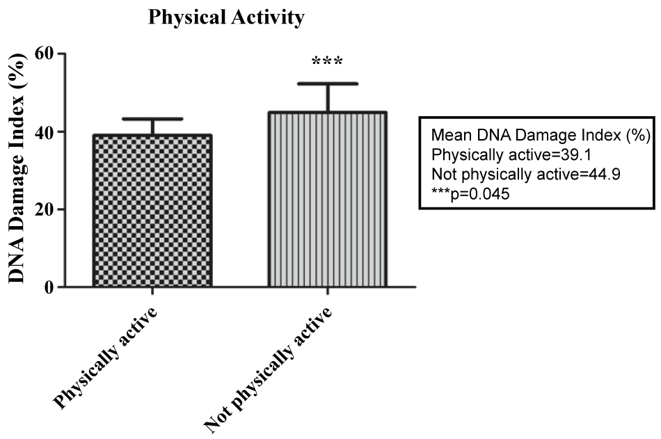
When comparing the average DDI (%) among the various qualitative variables (medicines in use, marital status, household income, race, occupation, education level, physical activity, classification of nutritional parameters and anti-DNA reagent), a significant difference (p = 0.045) was detected among those who practiced physical exercise (mean = 39.1% DDI) and those who did not (mean = 44.9% DDI), as shown in figure 1. Another significant difference (p = 0.006) was found between vitamin D users (mean = 37.2% DDI) and nonusers (mean = 45.3% DDI), according to figure 2. However, there was no statistical significance between the mean hs-CRP among the same qualitative variables.
.
Figure 1: DNA damage index in SLE patients physically active and not physically active.
View Figure 1
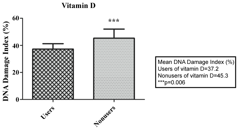
A multiple regression analysis model was used to check whether the independent variables: occupation, family income, education, physical activity, azathioprine, prednisone, chloroquine, folic acid and vitamin D explain the variable DDI. As the only independent variables that were statistically significant were physical activity and use of vitamin D, these were examined to determine if one had influence over the other with regard to DDI, and no such interference was found. However, these two independent variables explain 38.53% of the total variation of DDI. Thus, the patients in this study that were not physically active showed DDI 5.71% higher than the practitioners. Furthermore, patients using no vitamin D showed DDI 7.97% higher than users. The other independent variables were not statistically significant and therefore were removed from the model.
The anthropometric parameters, DDI (%), inflammatory markers and lipid profile of patients with SLE before and after treatment after completion of the crossover study are showed in table 2. The data were analyzed considering the effect of the patient as random. This test showed no significant effect of the pequi oil treatment for almost any variable analyzed, except for hs-CRP (p = 0.0161), which presented a statistically significant reduction compared to the placebo. For a 5% significance level a power of post hoc test for DDI was estimated to be 0.9887; for WC/HC of 0.9999 and the hs-CRP of 1.0000 to detect clinically important differences between treatments (pequi oil and placebo).
![]()
Table 2: The effect of pequi oil compared to placebo in patients with SLE in several variables.
View Table 2
Discussion
An inverse correlation was detected between DDI (%) and HDL levels in this study, also reported in other studies. One study compared individuals with normal TC, LDL, triglycerides and glucose, but with lowered HDL normolipidemic with controls, and observed that the former had high oxidative stress (measured by the plasma 8-isoprostanes) [35]. Plasma HDL has potent antioxidant activity so that synergy may occur in the inactivation of LDL oxidized lipids by enzymatic and non-enzymatic mechanisms [36]. And yet an inverse correlation between BMI and years of study was demonstrated, which corroborates the literature and means a higher BMI in people with lower educational levels [37,38]. It has been suggested that this association exists because the environmental conditions associated with low levels of education limit the knowledge of nutritious food and safe means for physical exercise, which causes metabolic deregulation and greater accumulation of calories [37].
Another important finding was the significant difference in the DDI between physical activity practitioners and non-practitioners observed in this study. Regular physical activity, besides functioning as an antioxidant to increase the expression of antioxidant enzymes, also induces a systemic increase in many cytokines with anti-inflammatory properties that protect against chronic disorders associated with low-grade systemic inflammation [39-42]. Therefore, results also confirm the protective effect of physical activity against oxidative stress. It is well documented in the literature that regular exercise results in adaptations in the antioxidant capacity protecting cells against the deleterious effects of oxidative stress and preventing subsequent cell damage [36]. It has been proven that physical activity is associated with a reduction in markers of oxidative stress in rodents [43], has a modulating effect on the antioxidant balance in women [44], increases total antioxidant capacity and reduces waist circumference in obese women [45]. Besides, regular exercise may stimulate DNA repair in healthy adults and the elderly, probably due an oxygen radical scavenger enzyme or repair enzyme mechanism [46]. Moderate exercise training is effective in increasing the endogenous antioxidant defenses and in reducing oxidative DNA damage in type 2 diabetes patients [47]. However, the current study is the first to demonstrate this relationship in SLE patients.
Our study also demonstrated that SLE patients using vitamin D had DDI (%) that was significantly lower than non-users. This information indicates that supplementation with vitamin D could reduce oxidative stress in these patients. This fact has also been suggested in a study conducted with type 2 diabetic black Americans [48]. Another study evaluated the status of vitamin D and markers of oxidative stress in older people who had compromised glucose metabolism and obtained an inverse association between these two parameters [49], which shows an antioxidant effect of this vitamin. There are no reports of clinical studies in the literature showing the relationship with SLE, but the protective effect of vitamin D against oxidative stress in these patients has been reported [50]. Multiple regression analysis with qualitative independent variables and the DDI shows that only physical activity and vitamin D reduced DNA damage.
Patients with SLE have an imbalance in the oxidant/anti-oxidant system independent of disease activity. In a previous study we demonstrated by comet assay that DDI increased in SLE patients when compared with healthy individuals, supporting an association between oxidative stress and SLE [19]. So, the present study analyzed if pequi oil in capsule, a rich antioxidant supplementation, has an antioxidant effect on patients with SLE, by means of comet assay. This method is one of many ways of measuring oxidative stress, and it can be applied in supplementation trials [35]. In this context, although pequi oil did not significantly reduce the DDI of the total group, it did significantly reduce hs-CRP.
First, this non-reduction of DDI after treatment runs contrary to the previous study with athletes of both genders who daily took 400 mg of pequi oil capsule for 14 consecutive days. These athletes showed decreased lipid peroxidation tested by TBARS, besides reduced DDI by comet assay. A reduction in muscle damage based on plasma evaluation of AST and ALT was also detected, the latter especially in women [26], probably due to the antioxidant activity of pequi [27]. This same study found a significant drop in TC and LDL in the group over 45 years, mostly in men [27]. In contrast to this finding, there was no significant reduction of TC, LDL, AST and ALT in the current crossover study. These controversial results, despite the similarity in the daily dose of pequi oil and higher total dose (60 days), might be explained by different methods of oil extraction in the two studies. In athletes, the extraction used the organic solvent chloroform, while the current study was by cold mechanical extraction. It has been shown that the extract has a higher mechanical peroxide content, demonstrating the greater vulnerability and lower content of carotenoids when compared to organic extracts [51]. Moreover, the storage time of the capsules with pequi oil was much higher in the current study than in athletes, which may have contributed to a further deterioration of carotenoids. Another factor that may have contributed to the reduction of this antioxidant was sterilization by radiation for the capsules used in the present study.
Secondly, even with the differences between the extraction methods of pequi oil, there was a significant reduction in hs-CRP in this work after pequi oil treatment, a result that confirms the previous study with athletes [26] in this aspect and also other studies evaluating the antioxidant intervention in inflammation by measuring hs-CRP [52,53]. The positive correlation between ROS and CRP has been confirmed in asymptomatic individuals [54] and also in some SLE patients [9], but not in all [10]. However, it seems that the interrelationship between inflammation-mediated CRP and certain forms of oxidative stress depended on the methods used. Moreover, it has been found that the CRP did not induce forms of DNA damage, single strand breaks and double strand breaks, but it was noted that prolonged exposure to CRP could have caused these types of damage [55]. This may explain the significantly reduced CRP values of the patients in our research after treatment with pequi oil, but not enough to reflect a reduction in damage caused by single and double strand breaks in DNA. Perhaps if the patients had had a higher initial value of CRP or if the treatment time were longer, a reduction in the DDI would have been observed beyond CRP reduction after treatment. A possible explanation for this finding, which is not balanced in the current crossover study, was the inclusion of patients with SLEDAI < 10; in other words, there were patients with high disease activity. An average level (3 mg/L) of CRP was recorded before beginning the use of the capsules, and the minimum (0.9 mg/L) and maximum (15 mg/L) and standard deviation (3.2 mg/L) values are within the classification proposed by Hooten et al. [55] to measure low and medium CRP.
High serum levels of hs-CRP are induced by various inflammatory conditions besides atherosclerosis. Thus, hs-CRP is not only a systemic inflammatory marker but also a local pro-atherosclerotic factor. Once a high level of hs-CRP is established, there is an increased cardiovascular risk [53]. A recent study even suggests that hs-CRP is a surrogate marker for cardiovascular risk in SLE patients, but this must be viewed with care because their values fluctuate with disease activity and infections [56]. Atherosclerosis is a well-known risk in SLE [57]. Moreover, the prevalence of and mortality from cardiovascular diseases is increasing among patients with SLE [58], and so these individuals have a greatly increased cardiovascular risk compared to the general population [59]. Therefore, the findings of the current study gain importance in clinical practice because they show a significant reduction in an inflammatory marker and predictor of atherosclerosis using antioxidant therapy.
At the same time, no connection between hs-CRP and the various qualitative and quantitative variables described here was noted, in contrast to the literature, which found an average increase in hs-CRP in SLE patients with higher BMI and among those that were less educated, had lower family income and higher disease activity [58]. The BMI in the study by Lee et al. [58] showed the highest correlation with hs-CRP among all variables. Another study demonstrated that both pre-obese and obese people in the general American population were more likely to have high levels of CRP than the normal weight controls [60]. Perhaps this same relationship was not detected in the current study, due to small sample size.
Despite having performed a sophisticated clinical trial (randomized double-blind crossover study), the current study has some limitations. There is no assurance that the patients properly ingested the capsules as directed, 1 capsule (400 mg of pequi oil) per day for 60 consecutive days. Another important factor was the difficulty for some patients to access the Clinical Laboratory at HUB. Ideally, the collection of blood and urine and anthropometric measurements were to take place on the 61st day after treatment or placebo. However, due to this limited access, sometimes the collection was carried out after completion of the therapy using capsules. Moreover, the extraction method used in this work was inferior to that used in a previous study, which may have affected the results. The peculiar and specific characteristics of the sample prevent the extrapolation of results to other populations of patients with SLE.
Conclusions
Although pequi oil did not significantly reduce the DNA damage index, it effectively reduced hs-CRP levels in these patients, indicating reduced inflammation with the use of antioxidant supplementation. This is important in clinical practice, in showing a significant reduction in an inflammatory marker that may predict atherosclerosis. Furthermore, the practice of physical activity and the use of vitamin D in patients with SLE could have an independent antioxidant effect.
Acknowledgement
The authors gratefully acknowledge the patients who participated in this research; University Hospital of Brasília, Farmacotécnica and Sabin Institute/Sabin Laboratories for technical support; and the National Council for Technological and Scientific Development (CNPq) for financial support.
References
-
Patavino T, Brady DM (2001) Natural medicine and nutritional therapy as an alternative treatment in systemic lupus erythematosus. Altern Med Rev 6: 460-471.
-
Robbins S, Cotran R, Kumar V (2000) Patologia estrutural e funcional. 5a Edição. Rio de Janeiro: Guanabara Koogan 1277.
-
Borba E, Latorre L, Brenol J, Kayser C, Silva N, et al (2008) Consenso de lúpus eritematoso sistêmico. Rev Bras Reumatol 48: 196-207.
-
Pons-Estel GJ, Alarcón GS, Scofield L, Reinlib L, Cooper GS (2010) Understanding the epidemiology and progression of systemic lupus erythematosus. Semin Arthritis Rheum 39: 257-268.
-
Bosch X (2011) Systemic lupus erythematosus and the neutrophil. N Engl J Med 365: 758-760.
-
Doria A, Arienti S, Rampudda M, Canova M, Tonon M, et al. (2008) Preventive strategies in systemic lupus erythematosus. Autoimmun Rev 7: 192-197.
-
Vasconcelos S, Goulart M, Moura J, Benfato V, Kubota L (2007) Espécies reativas de oxigênio de nitrogênio, antioxidantes e marcadores de dano oxidativo em sangue humano: principais métodos analíticos para sua determinação. Química Nova 30: 1323-1338.
-
Hassan S, Gheita T, Kenawy S, Fahim A, El-sorougy I, Abdou M (2011) Oxidative stress in systemic lupus erythematosus and rheumatoid arthritis patients: relationship to disease manifestations and activity. Int J Rheum Dis 14: 325-331.
-
Lozovoy MA, Simão AN, Panis C, Rotter MA, Reiche EM, et al. (2011) Oxidative stress is associated with liver damage, inflammatory status, and corticosteroid therapy in patients with systemic lupus erythematosus. Lupus 20: 1250-1259.
-
Avalos I, Chung CP, Oeser A, Milne GL, Morrow JD, et al. (2007) Oxidative stress in systemic lupus erythematosus: relationship to disease activity and symptoms. Lupus 16: 195-200.
-
Nuttall SL, Heaton S, Piper MK, Martin U, Gordon C (2003) Cardiovascular risk in systemic lupus erythematosus-evidence of increased oxidative stress and dyslipidaemia. Rheumatology (Oxford) 42: 758-762.
-
Mathis KW, Venegas-Pont M, Masterson CW, Stewart NJ, Wasson KL, et al. (2012) Oxidative stress promotes hypertension and albuminuria during the autoimmune disease systemic lupus erythematosus. Hypertension 59: 673-679.
-
Szeto YT, Benzie IF, Collins AR, Choi SW, Cheng CY, et al. (2005) A buccal cell model comet assay: development and evaluation for human biomonitoring and nutritional studies. Mutat Res 578: 371-381.
-
Vivek Kumar PR, Cheriyan VD, Seshadri M (2009) Could a strong alkali deproteinization replace the standard lysis step in alkaline single cell gel electrophoresis (comet) assay (pH > 13)? Mutat Res 678: 65-70.
-
Fairbairn DW, Olive PL, O'Neill KL (1995) The comet assay: a comprehensive review. Mutat Res 339: 37-59.
-
Dusinska M, Collins AR (2008) The comet assay in human biomonitoring: gene-environment interactions. Mutagenesis 23: 191-205.
-
McKelvey-Martin VJ, Green MH, Schmezer P, Pool-Zobel BL, De Méo MP, et al. (1993) The single cell gel electrophoresis assay (comet assay): a European review. Mutat Res 288: 47-63.
-
Cemeli E, Baumgartner A, Anderson D (2009) Antioxidants and the Comet assay. Mutat Res 681: 51-67.
-
Montalvão T, Miranda-vilela A, Roll M, Grisolia C, Santos-neto L (2012) DNA damage levels in systemic lupus erythematosus patients with low disease activity: An evaluation by comet assay. Adv Biosci Biotechnol 3: 983-988.
-
Hsieh CC, Lin BF (2011) Dietary factors regulate cytokines in murine models of systemic lupus erythematosus. Autoimmun Rev 11: 22-27.
-
Klack K, Bonfa E, Borba Neto EF (2012) Diet and nutritional aspects in systemic lupus erythematosus. Rev Bras Reumatol 52: 384-408.
-
Brown AC (2000) Lupus erythematosus and nutrition: a review of the literature. J Ren Nutr 10: 170-183.
-
Alves C, Resende J, Cruvinel R, Prado M (2008) Estabilidade da microestrutura e do teor de carotenoids de pós obtidos da polpa de pequi (Caryocar brasiliense Camb) liofilizada. Ciênc Tecnol Aliment 28: 830-839.
-
Miranda-vilela A, Grisolia C, Resck I, Mendonça M (2009) Characterization of the major nutritional components of Caryocar brasiliense fruit pulp by NMR spectroscopy. Quim. Nova 32: 2310-2313.
-
Miranda-vilela A, Grisolia C, Longo J, Peixoto R, De Almeida M, et al. (2014) Oil rich in carotenoids instead of vitamins C and E as a better option to reduce doxorubicin-induced damage to normal cells of Ehrlich tumor-bearing mice: hematological, toxicological and histopathological evaluations. J Nutr Biochem 25: 1161-1176.
-
Miranda-Vilela AL, Akimoto AK, Alves PC, Pereira LC, Gonçalves CA, et al. (2009a) Dietary carotenoid-rich pequi oil reduces plasma lipid peroxidation and DNA damage in runners and evidence for an association with MnSOD genetic variant – Val9Ala. Genet Mol Res 8: 1481-1495.
-
Miranda-Vilela AL, Pereira LC, Gonçalves CA, Grisolia CK (2009) Pequi fruit (Caryocar brasiliense Camb.) pulp oil reduces exercise-induced inflammatory makers and blood pressure of male and female runners. Nutr Res 29: 850-858.
-
Hochberg MC (1997) Updating the American College of Rheumatology revised criteria for the classification of systemic lupus erythematosus. Arthritis Rheum 40: 1725.
-
Nossent JC (1998). SLICC/ACR Damage índex in afro-caribbean patients with systemic lupus erythematosus: changes in and relationship to disease activity, corticosteroid therapy, and prognosis. J Rheumatol 25: 654-659.
-
Poirier P, Alpert M, Fleisher L, Thompson P, Sugerman H, et al. (2009) Cardiovascular evaluation and management of severely obese patients undergoing surgery: a science advisory from the American Heart Association. Circulation 120: 86-95.
-
Petri M, Orbai AM, Alarcón GS, Gordon C, Merrill JT (2012) Derivation and validation of the Systemic Lupus International Collaborating Clinics classification criteria for systemic lupus erythematosus. Arthritis Rheum 64: 2677-2686.
-
Engelman HM, Alekel DL, Hanson LN, Kanthasamy AG, Reddy MB (2005) Blood lipid and oxidative stress responses to soy protein with isoflavones and phytic acid in postmenopausal women. Am J Clin Nutr 81: 590-596.
-
Blackburn GL, Thornton PA (1979) Nutritional assessment of the hospitalized patient. Medical Clinics of North America, Philadelphia 14: 1102-1108.
-
Cornier MA, Després JP, Davis N, Grossniklaus DA, Klein S, et al. (2011) Assessing adiposity: a scientific statement from the American Heart Association. Circulation 124: 1996-2019.
-
Kontush A, De Faria E, Chantepie S, Chapman M (2005) A normotriglyceridemic, low HDL-cholesterol phenotype is characterised by elevated oxidative stress and HDL particles with attenuated antioxidative activity. Atherosclerosis 182: 277-285.
-
Kontush A, Chantepie S, Chapman MJ (2003) Small, dense HDL particles exert potent protection of atherogenic LDL against oxidative stress. Arterioscler Thromb Vasc Biol 23: 1881-1888.
-
Johnson W, Kyvik KO, Skytthe A, Deary IJ, Sørensen TI (2011) Education modifies genetic and environmental influences on BMI. PLoS One 6: e16290.
-
Sanchez-Vaznaugh EV, Kawachi I, Subramanian SV, Sánchez BN, Acevedo-Garcia D (2008) Differential effect of birthplace and length of residence on body mass index (BMI) by education, gender and race/ethnicity. Soc Sci Med 67: 1300-1310.
-
Colombini A, Lombardo G, Banfi G, Arpesella M, Pelissero G (2011) Athleticogenomics and elite athletes: a review of the state of the art and a possible relationship with inflammatory response. Ital J Public Health 8: 275-285.
-
Florencio GL, Gonçalves AK, Canário AC, Silva MJ (2011) Aging: a reflection about physical activity and oxidative stress in women. Acta Med Port 24 Suppl 4: 983-988.
-
Gomez-Cabrera MC, Domenech E, Viña J (2008) Moderate exercise is an antioxidant: upregulation of antioxidant genes by training. Free Radic Biol Med 44: 126-131.
-
Miranda-Vilela AL, Akimoto AK, Lordelo GS, Pereira LC, Grisolia CK, et al. (2012) Creatine kinase MM TaqI and methylenetetrahydrofolate reductase C677T and A1298C gene polymorphisms influence exercise-induced C-reactive protein levels. Eur J Appl Physiol 112: 183-192.
-
Gozal D, Nair D, Goldbart AD (2010) Physical activity attenuates intermittent hypoxia-induced spatial learning deficits and oxidative stress. Am J Respir Crit Care Med 182: 104-112.
-
Covas MI, Elosua R, Fitó M, Alcántara M, Coca L, et al. (2002) Relationship between physical activity and oxidative stress biomarkers in women. Med Sci Sports Exerc 34: 814-819.
-
Hen K, Bogdanski P, Szulińska M, Jabłecka A, Pupek-Musialik D (2010) [Influence of regular physical activity on oxidative stress in women with simple obesity]. Pol Merkur Lekarski 28: 284-288.
-
Cash SW, Beresford SA, Vaughan TL, Heagerty PJ, Bernstein L, et al. (2014) Recent physical activity in relation to DNA damage and repair using the comet assay. J Phys Act Health 11: 770-776.
-
Pittaluga M, Sgadari A, Dimauro I, Tavazzi B, Parisi P, et al. (2015) Physical Exercise and Redox Balance in Type 2 Diabetics: Effects of Moderate Training on Biomarkers of Oxidative Stress and DNA Damage Evaluated through Comet Assay. Oxid Med Cell Longev.
-
Jain SK, Manna P, Micinski D, Lieblong BJ, Kahlon G, et al. (2013) In African americans type 2 diabetic patients, is vitamin D deficiency associated with lower blood levels of hydrogen sulfide and cyclic adenosine monophosphate, and elevated oxidative stress? Antioxid Redox Signal 18: 1154-1158.
-
Gradinaru D, Borsa C, Ionescu C, Margina D, Prada GI, et al. (2012) Vitamin D status and oxidative stress markers in the elderly with impaired fasting glucose and type 2 diabetes mellitus. Aging Clin Exp Res 24: 595-602.
-
vinh quốc Luong K, Nguyên LT (2012) The beneficial role of vitamin D in systemic lupus erythematosus (SLE). Clin Rheumatol 31: 1423-1435.
-
Ribeiro M, Volas boas E, Riul T, Pantoja L, Marinho H, Santos A (2012) Influence of the extraction method and storage time on the physicochemical properties and carotenoid levels of pequi (Caryocar brasiliense Camb.) oil. Ciênc Tecnol Aliment Campinas.
-
Brighenti F, Valtueña S, Pellegrini N, Ardigo D, Rio D, et al. (2005) Total antioxidante capacity of the diet inversely and independently related to plasma concentration of high-sensitivity C-reactive protein in adult Italian subjects. Br J Nutr 93: 619-625.
-
Montecucco F, Mach F (2008) New evidences for C-reactive protein (CRP) deposits in the arterial intima as a cardiovascular risk factor. Clin Interv Aging 3: 341-349.
-
Kotani K, Taniguchi N (2012) Correlation Between High-sensitivity C-reactive Protein and Reactive Oxygen Metabolites During A One-year Period Among Asymptomatic Subjects. J Clin Med Res 4: 52-55.
-
Noren Hooten N, Ejiogu N, Zonderman AB, Evans MK (2012) Association of oxidative DNA damage and C-reactive protein in women at risk for cardiovascular disease. Arterioscler Thromb Vasc Biol 32: 2776-2784.
-
Mok CC, Birmingham DJ, Ho LY, Hebert LA, Rovin BH (2013) High-sensitivity C-reactive protein, disease activity, and cardiovascular risk factors in systemic lupus erythematosus. Arthritis Care Res (Hoboken) 65: 441-447.
-
Symmons DP, Gabriel SE (2011) Epidemiology of CVD in rheumatic disease, with a focus on RA and SLE. Nat Rev Rheumatol 7: 399-408.
-
Lee SS, Singh S, Magder LS, Petri M (2008) Predictors of high sensitivity C-reactive protein levels in patients with systemic lupus erythematosus. Lupus 17: 114-123.
-
Santos MJ, Carmona-Fernandes D, Canhão H, Canas da Silva J, Fonseca JE, et al. (2012) Early vascular alterations in SLE and RA patients-a step towards understanding the associated cardiovascular risk. PLoS One 7: e44668.
-
Visser M, Bouter LM, McQuillan GM, Wener MH, Harris TB (1999) Elevated C-reactive protein levels in overweight and obese adults. JAMA 282: 2131-2135.
