

International Journal of Diabetes and Clinical Research
Effect of Diabetes Mellitus on Metabolic Biomarker Levels in Patients with End Stage Renal Disease or Undergoing Total Joint Arthroplasty
J. Saluk1,2, Bansal V3, Banos A4, Hopkinson W4, Rees H4, Syed D2, Hoppensteadt D2, Abro S2,Iqbal O2 and Fareed J2*
1Stritch School o Medicine, Loyola University of Chicago, USA
2Hemostasis and Thrombosis, Department of Pathology, Loyola University Medical Center, USA
3Nephrology, Department of Internal Medicine, Loyola University Medical Center, USA
4Department of Orthopedic Surgery, Loyola University Medical Center, USA
*Corresponding author:
Fareed J, Hemostasis and Thrombosis Section, Department of Pathology, Loyola University Medical Center, 2160 S 1st Avenue, 60153, Maywood, IL, USA, E-mail: jfareed@luc.edu
Int J Diabetes Clin Res, IJDCR-2-027, (Volume 2, Issue 2), Research Article; ISSN: 2377-3634
Received: February 04, 2015 | Accepted: March 26, 2015 | Published: March 28, 2015
Citation: Saluk J, Bansal V, Banos A, Hopkinson W, Rees H, et al. (2015) Effect of Diabetes Mellitus on Metabolic Biomarker Levels in Patients with End Stage Renal Disease or Undergoing Total Joint Arthroplasty. Int J Diabetes Clin Res 2:027. 10.23937/2377-3634/1410027
Copyright: © 2015 Saluk J, et al. This is an open-access article distributed under the terms of the Creative Commons Attribution License, which permits unrestricted use, distribution, and reproduction in any medium, provided the original author and source are credited.
Abstract
Aim: Diabetes mellitus (DM) is a proven risk factor for diseases such as end stage renal disease (ESRD) and osteoarthritis (OA). Thus, it is not unreasonable to assume that some of the ESRD/OA pathogenesis, namely inflammation and metabolic dysfunction, could be attributed to DM as a comorbititiy. This study aims to investigate DM's effect on metabolic biomarker levels in patients with ESRD or undergoing total joint arthroplasty.
Methods: Under IRB approval, plasma samples were collected from 82 patients with ESRD prior to hemodialysis and from 39 patients prior to total hip and knee arthroplasty. Normal human plasma samples (female & male, 18-35 years old) were purchased from George King Biomedical Inc. (Overland Park, KS). Samples were stored at -80�C. Metabolic biochips were purchased from RANDOX (Co. Antrim, Northern Ireland) to test c-peptide, ferritin, IL-6, resistin, insulin, TNF-α, IL-1α, leptin and PAI-1. These biomarkers were tested in two batches: 82 ESRD and 17 normals, and 39 TJA and 25 normals.
Results & Conclusions: All biomarkers, except insulin, were significantly elevated in ESRD+DM compared to normal. For TJA+DM vs. normal, all biomarkers were increased except c-peptide and insulin, which were decreased, and ferritin, which showed no significant change. The only biomarker significantly increased in ESRD+DM compared to ESRD-DM, was leptin. Furthermore, ESRD+DM vs. normal were significant for leptin, but ESRD-DM vs. normal was not. This pattern is consistent with the leptin pattern seen in patients with ESRD+MetS. This suggests that elevated leptin levels seen in patients with ESRD may be due to DM and/or MetS, which are both highly prevalent in the ESRD population. For all other biomarkers, there was lack of significance in ESRD+DM vs. ESRD-DM, as well as in TJA+DM vs. TJA-DM. This suggests that the presence of DM may not substantially further alter the metabolic profiles seen in patients with ESRD or undergoing TJA.
Keywords
Diabetes mellitus, Metabolic syndrome, End stage renal disease, Total joint arthroplasty, Biomarkers, c-peptide, Ferritin, IL-6, Insulin, Resistin, TNF-α, IL-1α, Leptin, PAI-1
Introduction
Diabetes mellitus (DM) is a proven risk factor for diseases such as end stage renal disease (ESRD) and osteoarthritis (OA) [1-3]. Thus, it is not unreasonable to assume that some of the ESRD/OA pathogenesis, namely inflammation and metabolic dysfunction, could be attributed to DM as a comorbititiy. This study aims to investigate DM's effect on metabolic biomarker levels in patients with ESRD or undergoing total joint arthroplasty.
End stage renal disease
Chronic kidney disease, and its advancement to ESRD, has previously been linked to risk factors such as diabetes, hypertension and glomerulonephritis [1]. ESRD is marked by failure to remove waste products and uremia, requiring hemodialysis. Thus, an abnormal milieu of serum proteins and compounds, including metabolic waste products, would be expected in patients with ESRD. Whether or not part of the abnormal metabolic profile can be attributed to DM as a comorbitity or causative factor, is unclear.
Osteoarthritis and total joint arthroplasty
Osteoarthritis, a degenerative joint disease, is the leading cause of total joint arthroplasty (TJA) procedures in the United States [4,5]. Traditionally, OA has been seen as a degenerative joint disease due to long-term load-barring wear and tear of the joint. More recently, metabolic disregulation and inflammation have been indicated as contributing factors to the pathogenesis of OA and the need for TJA [2,6]. Diabetics have an increased risk for developing OA, possibly by accelerating joint damage through high serum levels of glucose and advanced glycosylation end products [2,3,7]. It has also been shown that low levels of insulin, as seen in patients with type 1 DM, decreases bone mineral density, and thus bone integrity [3,7,8]. In contrast, patients with type 2 DM have increased insulin and bone mineral density, but still have an increase risk of bone disorders, most likely attributed to dysregulation of bone and cartilage structure due to hyperglycemia and glycosylation [3,7,8]. While we expect to see altered metabolic biomarker patterns in patients undergoing TJA, we may also expect to see biomarker variance when comparing diabetic patients undergoing TJA to non-diabetic patients undergoing TJA.
Biomarkers
As insulin resistance and dysregulation are hallmarks of DM, we would expect to see altered insulin levels in ESRD/TJA patient populations. C-peptide is cleaved from pro-insulin and secreted with insulin, thus we would expect the c-peptide trend to mirror the changes in insulin9. However, it is important to note that c-peptide relies more on the kidney for clearance than insulin does, so we may expect to see higher levels of c-peptide than insulin in patients with ESRD [9].
Levels of leptin, an adipokine, are correlated to amount of adipose tissue, and at high levels has proinflammatory actions [10]. Nakhjavani et al. showed that leptin levels in patients with longstanding DM did not differ from normal, but that leptin was significantly lower in patients newly diagnosed with DM compared to normal and patients with longstanding DM [11]. Resistin and tissue necrosing factor alpha (TNF-α) are also secreted from inflamed adipose tissue and have been shown to increase in DM [10,12,13].
Ferritin is an iron storage protein best known for being increased during inflammation and infection, but is also linked to DM [14]. Plasminogen activator inhibitor 1 (PAI-1) prevents fibrin clot degradation and extracellular matrix turnover by inhibiting plasminogen activators, and thus plasmin [15]. Mechanisms involving PAI-1 may play a role in DM pathogenesis, thus we may expect to see increased PAI-1 in patients with DM [16,17].
Interleukin 6 (IL-6) is a pleiotropic cytokine that has both pro-inflammatory and anti-inflammatory actions. There have been numerous conflicting studies about IL-6 relation to and potential role in DM [16]. IL-6 can amplify the effects of other cytokines and possibly alter insulin secretion, thus it may contribute to the pathogenesis of DM, but is not likely a key player [17].
Interleukin 1 alpha (IL-1α) is another proinflammatory cytokine suggested to play a role in autoimmune processes [18]. Recently, it has been shown that fatty acids and metabolic stress can induce IL-1α release, triggering an inflammatory process in vasculature [18].
Materials and Methods
Patient recruitment
Patients diagnosed with ESRD and undergoing regular hemodialysis at Loyola University Ambulatory Dialysis Unit were recruited and informed consent was obtained. Additional inclusion criteria were age above 18 years, on hemodialysis for more than 90 days and free of any active infection or malignancy. Pre-dialysis blood was collected during the monthly laboratory blood collection at the dialysis unit. Patient sample collection was performed on the second dialysis of the week, prior to any heparin administration and before patients were connected to dialysis machine. All patient samples were drawn in non-fasting conditions.
Surplus plasma was obtained from patients scheduled to undergo TJA at Loyola University Medical Center. Plasma samples were collected during the routine pre-operative consultation and one day post-operative. All patient samples were drawn in non-fasting conditions.
Biomarker evaluation
Under IRB approval, plasma samples were collected from 82 patients with ESRD prior to hemodialysis and from 39 patients prior to total hip and knee arthroplasty (THA/TKA). Normal human plasma samples (female & male, 18-35 years old) were purchased from George King Biomedical Inc. (Overland Park, KS). Samples were stored at -80�C. Metabolic biochips were purchased from RANDOX (Co. Antrim, Northern Ireland) to test C peptide, ferritin, IL-6, resistin, insulin, TNF-α, IL-1α, leptin and PAI-1. These biomarkers were tested in two batches: 82 ESRD and 17 normals, and39 TJA and 25 normals.
The following information was collected from patients� electronic medical records: age, gender, BMI, HDL, triglycerides, systolic & diastolic BP, Hemoglobin A1C, fasting serum glucose, hypertension (HTN), DM, statin use and procedure type.
Statistical analysis
All data was analyzed using GraphPad Prism Software (San Diego, CA). Unpaired, non-parametric t-tests were conducted to determine the significance of patterns among variables.
Results
Biomarker evaluation in ESRD population
Population demographics and histograms of biomarker levels are shown in table 1 and figure 1. The following biomarkers were significantly elevated in patients with ESRD compared to normal: c-peptide, ferritin, IL-6, resistin, TNF-α, IL-1α, leptin, and PAI-1 as shown in table 2 and figure 2. No significance was found when comparing ESRD to normal for insulin. Leptin was significantly elevated in ESRD+DM, as compared to ESRD-DM. No significant difference was seen in other biomarkers for ESRD+DM vs. ESRD-DM.
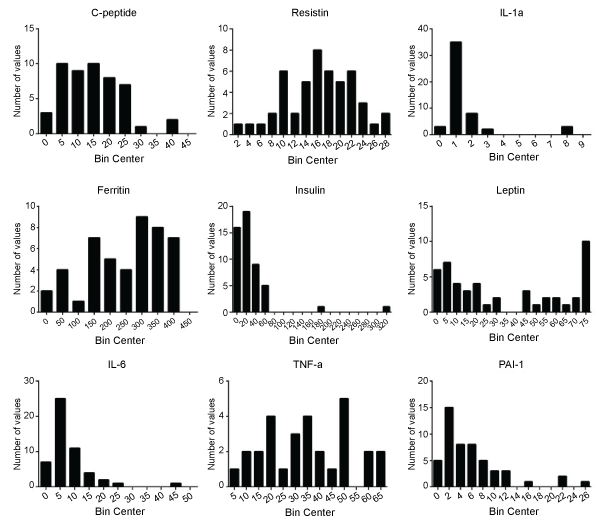
Figure 1: Frequency distribution of RANDOX biochip biomarker levels in patients with ESRD. Distributions vary across biomarkers for blood samples collected on
the 82 patients with ESRD. C-peptide, resistin and TNF-a show normal distributions. IL-6, insulin, IL-1α and PAI-1 show left skew, while leptin shows a bimodal
distribution. Bin centers created according to plasma sample biomarker concentrations: uIU/mL for insulin, pg/mL for IL-6, TNF-α and IL-1α, and ng/mL for
c-peptide, ferritin, resistin, leptin and PAI-1.
View Figure 1
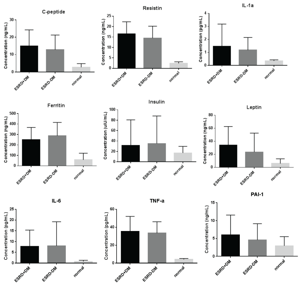
Figure 2: Bar graphs indicating biomarker means and standard deviations. The following biomarkers were significantly elevated in patients with ESRD compared to
normal: c-peptide (P< 0.0001), ferritin (P< 0.0001), IL-6 (P< 0.0001), resistin (P< 0.0001), TNF-α (P< 0.0001), IL-1α (P< 0.0001), leptin (P=0.01) and PAI-1 (P=0.05).
No significance was found when comparing ESRD to normal for insulin (P=0.4). Leptin was significantly elevated in ESRD+DM, as compared to ESRD-DM
(P=0.03). No significant difference was seen in other biomarkers for ESRD+DM vs. ESRD-DM.
View Figure 2
![]()
Table 1: Patient population demographics
View Table 1
![]()
Table 2: P-values for unpaired non-parametric t-tests run on biomarker data
View Table 2
Biomarker evaluation in TJA population
Population demographics and histograms of biomarker levels are shown in table 2 and figure 3. The following biomarkers were significantly elevated in patients undergoing TJA compared to normal: resistin, IL-1α, leptin, IL-6, TNF-α, and PAI-1 as shown in table 2 and figure 4. C-peptideand insulin was both decreased in TJA compared to normal. No significance was found when comparing TJA to normal for ferritin. For TJA+DM vs. TJA-DM, no significant difference was found in any biomarkers.
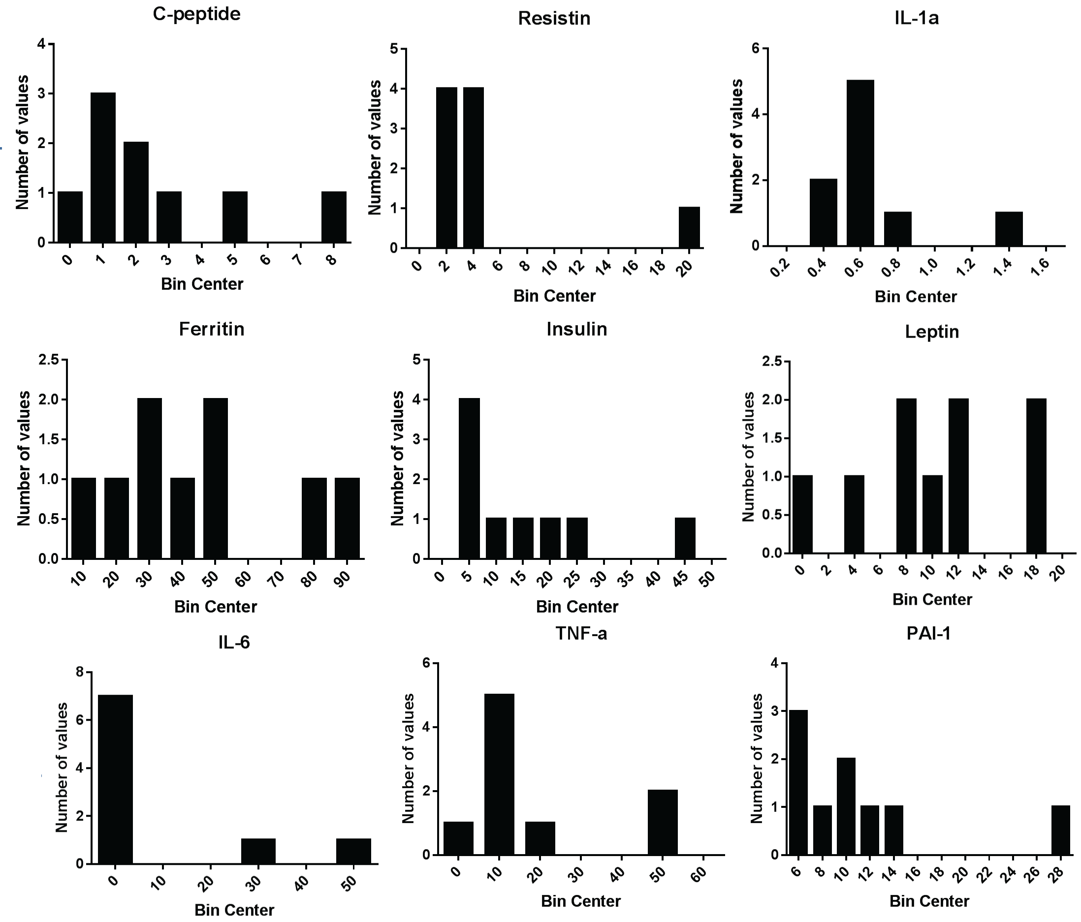
Figure 3: Frequency distribution of RANDOX biochip biomarker levels in patients undergoing TJA. Distributions vary across biomarkers for blood samples collected
on the 39 patients prior to TJA. Most had left skew trends, except for TNF-α and PAI-1, which showed more bimodal and uniform distributions, respectively. Bin
centers created according to plasma sample biomarker concentrations: uIU/mL for insulin, pg/mL for IL-6, TNF-α and IL-1α, and ng/mL for c-peptide, ferritin,
resistin, leptin and PAI-1.
View Figure 3
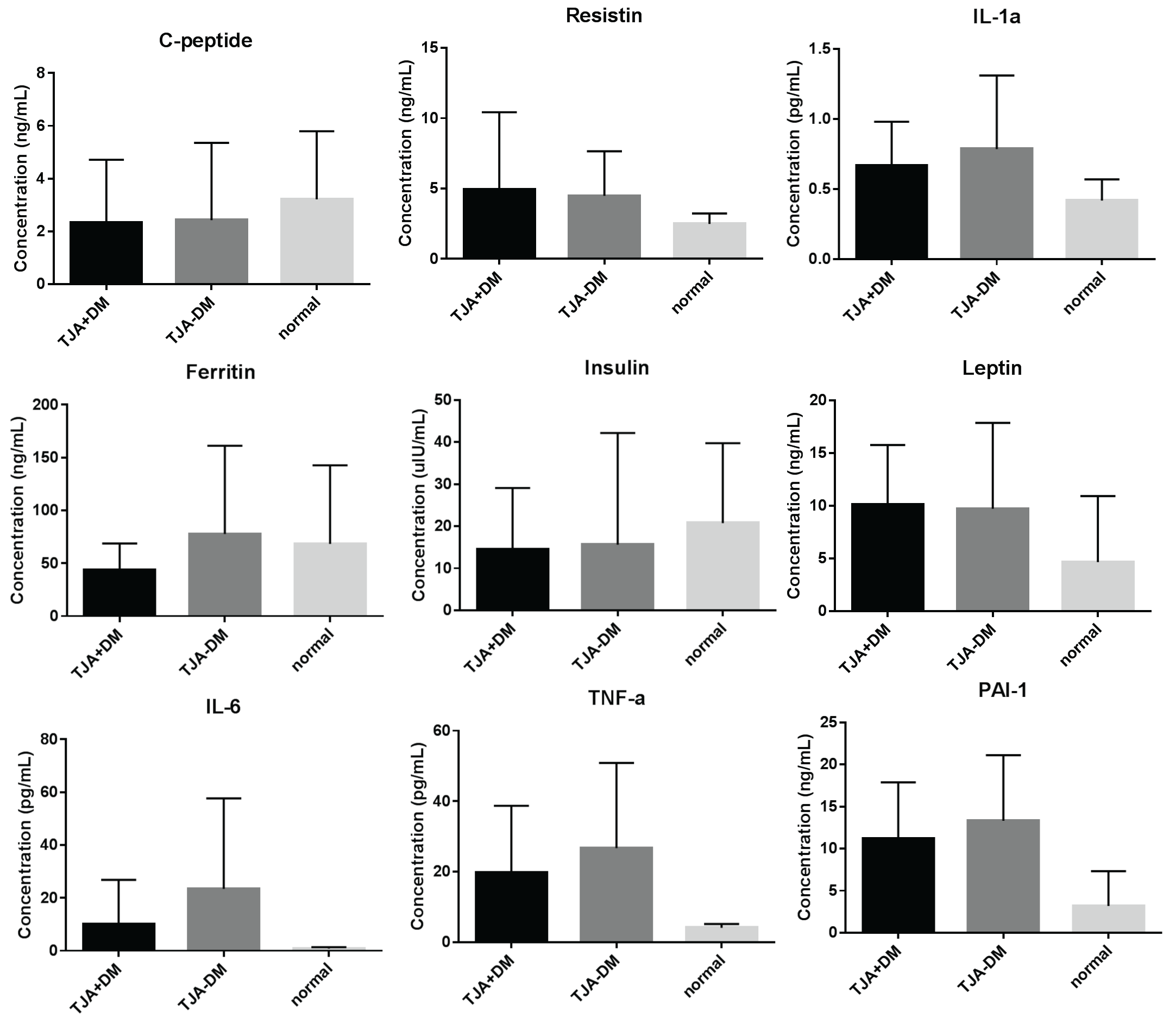
Figure 4: Bar graphs indicating biomarker means and standard deviations. The following biomarkers were significantly elevated in patients undergoing TJA
compared to normal: resistin (P=0.01), IL-1α (P=0.0003), leptin (P=0.01), IL-6 (P< 0.0001), TNF-α (P< 0.0001) and PAI-1 (P< 0.0001). C-peptide (P=0.01) and
insulin (P=0.01) were both decreased in TJA compared to normal. No significance was found when comparing TJA to normal for ferritin (P=0.6). For TJA+DM vs.
TJA-DM, no significant difference was found in any biomarkers.
View Figure 4
Discussion
Biomarker evaluation in ESRD population
Based on literature, we would expect to see increased levels of ferritin, PAI-1, resistin, IL1-α , TNF-α, c-peptide and insulin in patients with DM compared to normal, and no difference in IL-6 and leptin levels [11,12,14,15,17-21]. However, when investigating biomarker levels in patients with ESRD+DM vs. normal, we might expect to see a different metabolic profile than in patients with DM alone.
We have previously shown that c-peptide, resistin, IL-1α, ferritin, leptin, IL-6, PAI-1 and TNF-α are all increased in patients with ESRD compared to normal, while insulin is not [22]. Coupled with the fact that 60% of our ESRD population has DM, we expected and did see this same biomarker pattern in ESRD+DM vs. normal.
However, it was surprising to find that there was no significant difference between ESRD+DM and ESRD-DM, as we would have expected to see additional metabolic derangement in the diabetic group. The only biomarker that did show a significant increase in ESRD+DM compared to ESRD-DM, was leptin. Interestingly, we have previously shown that there was no correlation found between DM and biomarker levels in ESRD patients, but that DM was correlated to whether or not a patient fit the criteria for MetS. Thus it would make sense that the leptin pattern for DM mimics the leptin pattern for MetS in ESRD patients [22]. Here we see the same pattern of leptin levels in ESRD � DM as ESRD ± MetS: leptin is only significantly increased in ESRD patients compared to normal if they have DM (P< 0.0002), but is not significantly increased if they do not have DM (P=0.37).
Biomarker evaluation in TJA population
Although only 30% of the patients undergoing TJA had DM, biomarker patterns for TJA+DM vs. normal were consistent with TJA vs. normal biomaker patterns previously investigated. For TJA+DM vs. TJA-DM, no significant difference was found in any biomarkers, suggesting that the change in metabolic biomarker levels seen in patients undergoing TJA is due to joint disease processes, rather than due to DM as a comorbidity.
Limitations of this study
Many of these patients have numerous comorbidities, making it difficult to distinguish between biomarkers that are elevated as a result of the ESRD/OA/DM pathogenesis and biomarkers that are elevated as a result of comorbidities. Thus, more statistical analysis is required to investigate the correlations between biomarker levels and presence of various comorbidities.
As our patient populations all had either ESRD or joint deterioration, it was not possible to see whether patients with DM and no comorbidities (i.e. ESRD or OA) had elevated metabolic biomarkers as compared to baseline, as some literature suggested. It is possible that because patients with ESRD or undergoing TJA already have inflammatory processes and metabolic dysfunction occurring, the concurrence of DM did not substantially elevated biomarkers more than we would see in ESRD or TJA alone. However, if we looked at patients with DM but without ESRD, OA or other disease processes occurring, we may see a change in metabolic biomarker profile, when comparing DM to normal.
Appropriate controls for this study would have been matched for age, gender and BMI, to remove variations due to age-related disease processes, rather than ESRD, TJA, or DM disease states. However, recruitment of individuals above 60 years old who do not have any medical conditions is extremely slow, and thus normal samples (female & male, 18-35 years old) were purchased from George King Biomedical Inc. (Overland Park, KS). As shown in table 1, demographics (age, gender, BMI) are similar between ESRD+DM and ESRD-DM, and also between TJA+DM and TJA-DM, with the exception of the apparent age difference seen between TJA+DM and TJA-DM. While, this was not the focus of our study, a larger cohort would allow us to demonstrate any potential demographic differences.
We did not separate patients based on what type of DM they had, and as the two types do have different pathogenesis, we may expect to see some variation between the two groups. Thus, additional investigation of biomarker levels in type 1 DM and type 2 DM would be useful. It is also important to point out that the metabolic profile may vary from patient to patient depending on how far their disease has progressed.
Many of patients are treated with numerous anti-inflammatory, analgesic, antidiabetic and other medications. Some of these medications may have a modulatory effect on the generation of biomarkers. It would be difficult to differentiate the specific effects produced by antidiabetic medications alone from effects of other medications. It is also important to note that most of the ESRD+DM and TJA+DM patients are being treated with antidiabetic medications, which may affect the measured analyses. Upon review of the available HbA1C and glucose levels, there was a mix between pre-diabetic and non-diabetic patients in the non-DM groups. Moreover, it was not our objective to differentiate between pre-diabetic and non-diabetics.
Plasma samples drawn were from patients with ESRD as part of a maintenance dialysis clinic-based draw, which was completed in 3 shifts. Thus it was not possible to obtain fasting samples. Likewise, the arthroplasty plasma samples were obtained at a pre-surgical testing panel and thus can also be assumed non-fasting glucose samples. However, most of the metabolic biomarkers included in this investigation have relatively long half-lives and do not change prandially. It�s unlikely that fasting would have altered these results.
Some of the patients undergoing TJA had very little documented in their chart, possibly because Loyola University Medical Center is not their primary healthcare provider. Thus it is possible that some patients may have had DM that was not documented in the charts we had access to during this project.
Conclusions
The altered metabolic profiles seen in patients with ESRD or undergoing TJA, as compared to normal, suggests that there is metabolic dysfunction present in these disease states, which possibly contributes to the pathogenesis and overall clinical outcome in these groups. The only biomarker significantly increased in ESRD+DM compared to ESRD-DM, was leptin. Furthermore, ESRD+DM vs. normal were significant for leptin, but ESRD-DM vs. normal was not. This pattern is consistent with the leptin pattern seen in patients with MetS. This suggests that the elevated leptin levels seen in patients with ESRD may be due to DM and/or MetS, which are highly prevalent in the ESRD population. For all other biomarkers, there was lack of significance between ESRD+DM and ESRD-DM, as well as between TJA+DM and TJA-DM. This suggests that the presence of DM may not substantially further alter the metabolic profiles seen in patients with ESRD or undergoing TJA.
Acknowledgements
The authors gratefully acknowledge the assistance of the orthopedic surgery staff and dialysis center staff at the Loyola University Medical Center for the facilitation of the collection of the blood samples from patients undergoing total joint arthroplasty and dialysis, respectively. We are also thankful to the staff of hemostasis and thrombosis research laboratories for their skillful assistance in generating some of the information included in this manuscript. Funding is from institution.
References
-
Kabir MS, Dutta PK, Islam MN, Hasan MJ, Mondol G (2012) Prevalence of risk factors of chronic kidney disease in adults. Mymensingh Med J 21: 605-610.
-
Piva SR, Susko AM, Khoja SS, Josbeno DA, Fitzgerald GK, et al. (2015) Links between osteoarthritis and diabetes: implications for management from a physical activity perspective. Clin Geriatr Med 31: 67-87, viii.
-
Yan W, Li X (2013) Impact of diabetes and its treatments on skeletal diseases. Front Med 7: 81-90.
-
Martin GM, THornhill TS, Katz JN (2014) Total Knee Arthroplasty.
-
Erens GA, Thornhill TH, Katz JN (2014) Total hip arthroplasty.
-
Thijssen E, van Caam A1, van der Kraan PM2 (2014) Obesity and osteoarthritis, more than just wear and tear: pivotal roles for inflamed adipose tissue and dyslipidaemia in obesity-induced osteoarthritis. Rheumatology (Oxford) .
-
Hofbauer LC, Brueck CC, Singh SK, Dobnig H (2007) Osteoporosis in patients with diabetes mellitus. J Bone Miner Res 22: 1317-1328.
-
Piva SR, Susko AM, Khoja SS, Josbeno DA, Fitzgerald GK, et al. (2015) Links between osteoarthritis and diabetes: implications for management from a physical activity perspective. Clin Geriatr Med 31: 67-87, viii.
-
Henriksen JH, Tronier B, B�low JB (1987) Kinetics of circulating endogenous insulin, C-peptide, and proinsulin in fasting nondiabetic man. Metabolism 36: 463-468.
-
Sartori-Cintra AR, Aikawa P, Cintra DE (2014) Obesity versus osteoarthritis: beyond the mechanical overload. Einstein (Sao Paulo) 12: 374-379.
-
Nakhjavani M, Morteza A, Nargesi AA, Mostafavi E, Esteghamati A (2013) Appearance of leptin-HSP70 correlation, in type 2 diabetes. Meta Gene 1: 1-7.
-
Onuma H, Osawa H, Makino H (2008) [Role of resistin in insulin resistance]. Rinsho Byori 56: 698-704.
-
Ozer G, Teker Z, Cetiner S, Yilmaz M, Topaloglu AK, et al. (2003) Serum IL-1, IL-2, TNFalpha and INFgamma levels of patients with type 1 diabetes mellitus and their siblings. J Pediatr Endocrinol Metab 16: 203-210.
-
Sun L, Franco OH, Hu FB, Cai L, Yu Z, et al. (2008) Ferritin concentrations, metabolic syndrome, and type 2 diabetes in middle-aged and elderly chinese. J Clin Endocrinol Metab 93: 4690-4696.
-
Metabolic Syndrome Array 1. Randox Laboratories Limited. EV3755.
-
Kristiansen OP, Mandrup-Poulsen T (2005) Interleukin-6 and diabetes: the good, the bad, or the indifferent? Diabetes 54 Suppl 2: S114-124.
-
Longo PL, Artese HP, Rabelo MS, Kawamoto D, Foz AM, et al. (2014) Serum levels of inflammatory markers in type 2 diabetes patients with chronic periodontitis. J Appl Oral Sci 22: 103-108.
-
Garlanda C, Dinarello CA, Mantovani A (2013) The interleukin-1 family: back to the future. Immunity 39: 1003-1018.
-
Correia ML, Haynes WG (2006) A role for plasminogen activator inhibitor-1 in obesity: from pie to PAI? Arterioscler Thromb Vasc Biol 26: 2183-2185.
-
Walcher D, Babiak C, Poletek P, Rosenkranz S, Bach H, et al. (2006) C-Peptide induces vascular smooth muscle cell proliferation: involvement of SRC-kinase, phosphatidylinositol 3-kinase, and extracellular signal-regulated kinase 1/2. Circ Res 99: 1181-1187.
-
Lechleitner M, Koch T, Herold M, Dzien A, Hoppichler F (2000) Tumour necrosis factor-alpha plasma level in patients with type 1 diabetes mellitus and its association with glycaemic control and cardiovascular risk factors. J Intern Med 248: 67-76.
-
Saluk J, Bansal V, Hoppensteadt D, Syed D, Abro S, et al. (2014) Prevalence of metabolic syndrome in patients with end stage renal disease and relevance of biomarkers. Int Angiol .
