
 Neurosurgery Cases and Reviews is an international, peer reviewed, open access journal that deals with the medical specialty concerned with the prevention, diagnosis, surgical treatment, and rehabilitation of disorders affecting portions of the nervous system including the brain, spinal cord, peripheral nerves, and extra-cranial cerebrovascular system.
All the published articles are permanently archived without any restriction barriers to access under terms of creative commons attribution license. The submitted manuscripts undergo thorough double blind peer review process and are accepted only by recommendation of editorial experts. The journal invites various type of articles covering but not limited to ablative surgery, astroglias, brain hemorrhages, cervical spinal stenosis, cranial/peripheral nerve pain, deep brain stimulation surgery, drug-resistant epilepsy, glioblastomas, hydrocephalus infections, intracerebral hemorrhage, intractable psychiatric disorders, invasive stereotactic neurosurgery, lumbar spinal stenosis, movement disorders, moyamoya disease, skull fractures, spinal cord trauma, spinal disc herniation, traumatic injuries of peripheral nerves, tumors of the spine, spinal cord and peripheral nerves, vascular malformations, etc.
Journal Information
Title: Neurosurgery - Cases and Reviews
ISSN: 2643-4474
Editor-in-chief: Francesco Maiuri
NLM title abbreviation: Neurosurg Cases Rev
ISO abbreviation: Neurosurg Cases Rev
Other titles: NCR
Category: Neurosurgery/ Medicine
DOI: 10.23937/2643-4474
Peer review: Double blind
Review speed: 3 weeks
Fast-track review: 10 days
Publication format (s): Electronic and print
Publication policy: Open Access; COPE guide
Publication type(s): Periodicals
Publisher: ClinMed International Library
Country of publication: USA
Language: English
Contact email: contact@clinmedjournals.org
Articles Search by Keyword | Journal title | Author name | DOI
Open Access DOI:10.23937/2643-4474/1710178 Haiyue Jin, Alexander M. Lopez, Michael Y. Oh, Yarah M. Haidar, Xiao-Tang Kong, Jerica Lomax, Aaron Simon, William Yong and Hansen Bow Article Type: Case Report and Literature Review | First Published: January 21, 2026 The 56-year-old presented with progressive left upper and lower extremity weakness. He underwent resection of the ventral intradural lesion at C6-7. The surgery involved a C5-7 corpectomy for resection and a C2- T3 posterior spinal fusion for stabilization. His weakness resolved postoperatively. We then present a systematic review of the literature on the dissemination of low-grade posterior fossa ependymoma. Fifteen studies were identified, involving 23 patients....
| |
Open Access DOI:10.23937/2643-4474/1710176 Thoracic Spinal Lymphangioma in an Adult: A Case Report Salcedo-Hernandez Missael de Jesús, MD, Martinez-Nava Alexis Oziel, MD, Cobián-Estrada Yonatan Emmanuel, MD, Ramos-Sánchez Gilberto Daniel, MD, Franco-Del Toro Perla Guadalupe, MD, Plazola-Valle Citlalli Xmell, MD and Andrade-Ramos Miguel Ángel, MD Article Type: Case Report | First Published: January 18, 2026 This case involves a 56-year-old female patient presented to the clinic with complaints of persistent cervical pain and predominant weakness in the lower extremities over the past three months. Physical examination revealed clinical signs at the dorsal level. Further imaging revealing a lesion within the spinal canal exerting a mass effect on neural structures. An elective procedure was performed, including a fluoroscopy-guided laminectomy at the previously identified affected level (T2), the le...
| |
Open Access DOI:10.23937/2643-4474/1710175 Acute Deterioration in a Pediatric Patient with a Transitional Lipoma - A Case Report Gabrielle Gour-Provençal, MD, FRCPC, MSc1,2, Timothée De Saint-Denis, MD3, Pauline Lallemant-Dudek, MD1 and Hina Simonnet, MD Article Type: Case Report | First Published: January 18, 2026 Lumbosacral lipomas, the most common form of spinal dysraphism, represent a challenge in clinical decision making as optimal management in asymptomatic patients is still unclear. There is ongoing debate whether these patients should undergo prophylactic surgery or not. Especially when multiple management options exist, shared decision making should be prioritised as it has been shown to decrease decisional conflict and anxiety and increase decisional satisfaction (despite outcome), knowledge, qu...
| |
Open Access DOI:10.23937/2643-4474/1710174 Tiago Oliveira, Miguel Ferreira, Teresa Pinheiro, João Lourenço and Joaquim Monteiro Article Type: Case Report | First Published: January 17, 2026 Deep brain stimulation (DBS) is an established treatment for advanced Parkinson’s disease, and although generally safe, complications related to the implantable impulse generators (IPGs), such as infection, skin erosion, or device malfunction, can arise. Malignancy arising at the IPG implantation site is exceedingly rare, with few cases previously reported, related to pacemakers IPGs. We present case of invasive ductal breast carcinoma developing adjacent to a DBS IPG. A 77-year-old woman with...
| |
Open Access DOI:10.23937/2643-4474/1710173 Emna Elouni, Atef Ben Nsir, Asma Elmir, Lobna Mahjoub and Wiem Boudabbous Article Type: Case Report | First Published: September 08, 2025 We report the case of a 27-year-old male with a history of ventriculoperitoneal shunt placement at 8 months of age for congenital hydrocephalus. He presented to the emergency department two days after a minor cervical trauma, exhibiting signs of intracranial hypertension and a rapid decline in consciousness. On examination, his Glasgow Coma Scale score was 12/15. Cervical spine radiography revealed a disconnection of the shunt tubing at the cervical level....
| |
Open Access DOI:10.23937/2643-4474/1710172 Olfactory Groove Meningioma Presenting Solely with Visual Impairment: An Atypical Case Omar Abu Saadeh, Lena Mary Houlihan, Alan Beausang, Melanie Lang Orsini and DonnachaO' Brien Article Type: Case Report | First Published: August 09, 2025 Olfactory groove meningiomas (OGMs) are rare intracranial tumours that arise from the anterior cranial fossa, typically originating at the cribriform plate, planum sphenoidale, or fronto sphenoidal suture. Due to their slow-growing and insidious nature, OGMs often go undetected until they reach considerable size, making them among the largest central nervous system (CNS) tumours at diagnosis. Common presenting symptoms include headache, anosmia, personality changes, and visual impairment; howeve...
| |
Open Access DOI:10.23937/2643-4474/1710170 Djalma de Campos Goncalves Article Type: Case Report | First Published: May 16, 2025 Arachnoid cysts are benign congenital malformations typically located within the cerebrospinal fluid spaces of the brain. They are often asymptomatic and discovered incidentally. However, complications such as hemorrhage can render them clinically significant, especially following minor trauma. We present the case of a 19-year-old male who developed a progressive headache after mild head trauma. Neuroimaging revealed a right frontoparietal subacute subdural hematoma with significant midline shif...
| |
Open Access DOI:10.23937/2643-4474/1710169 Visual Prognostic Factors in Surgery of Tubercule Sellae Meningiomas Lotfi Boublata, Oumeima Bouzrara, Belgacem Boudjadja and Amine Boukhanoufa Article Type: Case Report and Literature Review | First Published: February 21, 2025 This is a retrospective study of patients operated on for TS meningioma, conducted in the Department of Neurosurgery at Dr. Benbadis University Hospital in Constantine, Algeria. All patients were evaluated by an ophthalmologist before and after surgery. MRI is used to study tumor size, tumor extension and tumor relationships with vessels of the anterior part of the polygon of Willis....
| |
Open Access DOI:10.23937/2643-4474/1710167 Intracranial Birth Injuries in the Full-Term Neonate: Case Report and Literature Review Dylan Calame, MD, Melanie Mandell, BA, Izaz Riaz, MD, Fabio Grassia, MD and Muhammad Riaz, MD Article Type: Case Report & Literature Review | First Published: February 14, 2025 Symptomatic traumatic epidural hematoma (EDH) in a newborn is extremely rare. These injuries can occur for various reasons, such as prolonged labor, fetal distress, assisted delivery techniques such as forceps or vacuum extractors, or cesarean sections....
| |
Open Access DOI:10.23937/2643-4474/1710166 Mikhail Shkarubo, Alexander Konovalov and Vladimir Tyurin Article Type: Case Report & Literature Review | First Published: 2024/12/14 Colloid cysts of the third ventricle are rare congenital benign lesions that can cause acute occlusion of CSF pathways with the risk of a fatal outcome. Up to 6% of colloid cysts are asymptomatic, incidental findings. Colloid cysts tend to slowly increase in size over a long period of time. Management of patients with colloid cysts of the third ventricle depends on clinical and radiological findings. Spontaneous shrinkage (rupture) of the colloidal cyst is an extremely rare phenomenon, described...
| |
Open Access DOI:10.23937/2643-4474/1710165 M. Colosimo, C. Thole, B. Sommer, M.C. Frühwald, A. Berlis and E. Shiban Article Type: Case Report | First Published: 2024/12/05 Spinal arteriovenous malformations (AVM) are rare vascular occurrences that, if left untreated, can result in devastating neurologic sequelae. Prevalence, especially in the pediatric population, is low but requires prompt diagnosis and management to avoid neurologic impairment. Embolization has been advocated as the treatment of choice for spinal AVMs. We present a rare case of a spinal AVM in an 11-year-old male managed with endovascular embolization during continuous intraoperative monitoring ...
| |
Open Access DOI:10.23937/2643-4474/1710164 Hidden Hazard of Remote Cerebellar Hemorrhage after Cervical Spine Surgery Barnabas Obeng-Gyasi, BS, Emma Stephens Love, BA, MS, Mugtaba Swar-Eldahab, MS, Matthew Blackwell, MS and Gordon Mao, MD Article Type: Case Report | First Published: 2024/10/30  Remote cerebellar hemorrhage (RCH) is a rare but potentially serious complication of spinal surgery, particularly after decompressive procedures and instrumented fusion. The complex interplay between cerebrospinal fluid leaks and hemorrhage risk necessitates a high index of suspicion and prompt management to optimize patient outcomes.... Remote cerebellar hemorrhage (RCH) is a rare but potentially serious complication of spinal surgery, particularly after decompressive procedures and instrumented fusion. The complex interplay between cerebrospinal fluid leaks and hemorrhage risk necessitates a high index of suspicion and prompt management to optimize patient outcomes....
| |
Open Access DOI:10.23937/2643-4474/1710163 Woven EndoBridge (WEB) Device used for Vertebral Artery Sacrifice Jason J. Sims, BS, Bhageeradh Mulpur, MD, Nick Khattar, MD and Adam A. Arthur, MD, MPH Article Type: Case Report | First Published: 2024/10/11 The decision to sacrifice a vessel in the context of endovascular interventions arises from specific clinical indications where alternative treatments pose either higher risks or are simply not feasible. One critical indication is in the control of uncontrolled hemorrhage, particularly in cases of severe vascular trauma or bleeding situations. Additionally, sacrificing a vessel may be considered for managing aneurysms that are difficult to repair surgically or with other endovascular methods, es...
| |
Open Access DOI:10.23937/2643-4474/1710162 Razan Almufarriji, MD and Abdullah Alobaid, MD, FRCSC Article Type: Case Report | First Published: 2024/07/06 Meningiomas encasing an aneurysm are considered one of the rare presentations encountered in neurosurgical services. To the best of our knowledge, this is the fourth case describing a frontotemporal meningioma encasing a middle cerebral artery aneurysm. Management of such cases has many aspects to consider and scenarios to be prepared for....
| |
Open Access DOI:10.23937/2643-4474/1710161 CNS Embryonal Tumour with Desmoplastic Reaction Treated with Ultra-NPT Bionanocatalysts Tessy Lopez-Goerne, Francisco J Padilla-Godínez, Daniel Alvárez-Amador, Antonela Gonzalez-Bondani, Rafael Valiente, Fernando Chico Ponce de Leon, Vicente Gonzalez-Carranza and José Omar Navarro-Fernández Article Type: Case Report | First Published: 2024/07/06 Central nervous system (CNS) embryonal tumors, lacking a distinct molecular signature and categorized as "Not Otherwise Specified (NOS)", are WHO grade IV neoplasms with significant genetic diversity. They typically exhibit immature cell sheets with round, oval, or angulated nuclei, often located supratentorially and characterized by a desmoplastic reaction. Diagnosis relies on MRI and molecular analysis, with traditional management involving surgery, radiotherapy, and chemotherapy. However, rec...
| |
Open Access DOI:10.23937/2643-4474/1710160 Hideki Nakajima, MD, Morio Takasaki, MD, Taku Hongo, MD, Katsuma Iwaki, MD, PhD, Ryota Miyake, MD and Yasuhiro Fujimoto, MD Article Type: Case Report | First Published: 2024/06/29 We report two cases of a usual saccular cerebral aneurysm with an iatrogenic lacerated aneurysmal neck during surgical clipping. In both cases, the aneurysmal neck was lacerated iatrogenically, and the aneurysm was clipped safely after suturing the lacerated portion. In treatment of a lacerated aneurysmal neck during surgical clipping of usual saccular cerebral aneurysms, the aneurysm may be considered to be clipped after suturing the lacerated portion. However, trapping and/or bypass surgery be...
| |
Open Access DOI:10.23937/2643-4474/1710159 Félix LeBlanc, Lyndon Boone, Timothy Noble, Jane Burns, Dhany Charest<, Maxime Richer and Antonios El Helou Article Type: Case Report | First Published: 2024/06/12 5-ALA is a prodrug used to selectively illuminate high-grade glioma (HGG) tissue intra-operatively and optimize resection. Here, we present some of the first preliminary results on resection rates, survival analysis, and adverse effects from a single Canadian center....
| |
Open Access DOI:10.23937/2643-4474/1710158 Emal Lesha, MD, L Erin Miller, BA, Taylor Orr, MS, Michael S Muhlbauer, MD, Manjari Pandey, MD, Brent A Orr, MD, PhD, Andrew B Boucher, MD and Sarada Wellikoff, MD Article Type: Case Report | First Published: 2024/04/17 Atypical teratoid/rhabdoid tumor (AT/RT) is a Grade 4 embryonal tumor, rare and aggressive, most often presenting in children 0-5 years of age. In the adult population, AT/RTs are exceedingly rare, and usually located intracranially in the supratentorial compartment. AT/RTs of the spine are even more uncommon. Here, we detail the case of a young adult with AT/RT of the lumbar spine who underwent surgical resection of the lesion followed by radiation and chemotherapy. We also provide a comprehens...
| |
Open Access DOI:10.23937/2643-4474/1710157 An Unusual Complication, Rupture of the Intraoperative Ultrasound Probe Sheath: A Case Report Erkin Ozgiray, Bilal Bahadır Akbulut and Mustafa Serdar Boluk Article Type: Case Report | First Published: 2024/03/15 Intraoperative ultrasound (USG) imaging is employed by many neurosurgical institutes for localizing lesions with better accuracy in real-time. It’s often used with sterilized probes or purpose-built sterile sheaths. It has also been reported to be used with a sterile latex glove filled with USG gel. We report a case of ruptured intraoperative USG sheath which was made out of a sterile latex glove causing contamination of the surgical field....
| |
Open Access DOI:10.23937/2643-4474/1710156 Persistent Primitive Trigeminal Artery Aneurysm Dantas Mageste Ferreira, MD, M.Sc, Lano de Sousa Moreira, MD, Taianne Fiore Schumann, MD, Jéssica Aguilar da Silva, Tarciano Costa Nascimento, MD, André Guimarães Soares, MD, Fernando Miguel Resck Gadbem, MD and Ítalo Guilherme Giarola de Freitas Mariano, MD Article Type: Case Report | First Published: 2024/03/14 The Persistent Primitive Trigeminal Artery (PPTA) is an example of persistent carotid-vertebrobasilar anastomoses, in which the basilar artery and its branches are primarily supplied by an anastomotic arterial branch originating from the cavernous segment of the Internal Carotid Artery (ICA). Such a structure constitutes the largest of fetal carotid-basilar anastomotic arteries and usually involutes after the development of the Posterior Communicating Artery (P-com)....
| |
Open Access DOI:10.23937/2643-4474/1710155 Rare Galen Vein Aneurysm in a 40-Year-Old Woman: In-Depth Analysis of an Exceptional Case Dantas Mageste Ferreira, MD, MSc, Vitor de Deus da Rocha Ribeiro Gonçalves, MD, Lano de Sousa Moreira, MD, Taianne Fiore Schumann, MD and Jéssica Aguilar da Silva Article Type: Case Report | First Published: 2024/02/09 Vein of Galen aneurysm is a rare vascular condition commonly diagnosed in children. This type of aneurysm arises from the persistence of the embryonic median prosencephalic vein, precursor to the Galen vein. This paper highlights the rarity of this phenomenon in a 40-year-old woman. The aim of this study is to demonstrate that conservative treatment of this malformation can be a valid option in oligosymptomatic cases without signs of hydrocephalus....
| |
Open Access DOI:10.23937/2643-4474/1710154 Recurrent Post-Traumatic Carotid-Cavernous Fistula after Endovascular Treatment Dantas Mageste Ferreira, MD, MSc, Jéssica Aguilar da Silva, Vitor de Deus da Rocha Ribeiro Gonçalves, MD, Taianne Fiore Schumann, MD and Lano Sousa Moreira, MD Article Type: Case Report | First Published: 2024/01/24 The High-Flow Type A Carotid-Cavernous Fistula (CCF) is the direct communication between the cavernous segment of the internal carotid artery (ICA) and the cavernous sinus. Its most frequent etiology is trauma, more commonly occurring in males. Clinically, it is characterized by the Dandy triad of hyperemia, exophthalmos, and ocular bruit. The described case involves a male patient who presented with a classic high-flow CCF, underwent coil embolization, showed a satisfactory response in the firs...
| |
Open Access DOI:10.23937/2643-4474/1710153 Intraoperative Monitoring of Carotid Endarterectomy with Fluorescein Coupled to Surgical Microscope Dantas Mageste Ferreira, MD, MSc, Marcos Dellaretti, MD, PhD1, Vitor de Deus da Rocha Ribeiro Gonçalves, MD, Lano de Sousa Moreira, MD, Taianne Fiore Schumann, MD and Jéssica Aguilar da Silva Article Type: Case Report | First Published: 2024/01/14 The article aims to present partial results from an ongoing clinical trial, including fourteen patients undergoing endarterectomy with an innovative technique. The male gender accounted for 71.4% of the sample, with a mean age of 67.93 (± 7.75) years for the patients. Among comorbidities, systemic arterial hypertension was the most prevalent at 85.7%, followed by dyslipidemia at 57.1%. ...
| |
Open Access DOI:10.23937/2643-4474/1710152 Titanium Interbody Cage: A Case Series Demonstrating Expandable Technology in Spinal Surgery in Peru Christian A Yataco-Wilcas, Luis A Lengua-Vega, Bruno E Diaz-Llanes, Yosimar S Coasaca-Tito and Cristian E Salazar-Campos Article Type: Descriptive Study | First Published: 2023/01/07 Lumbar interbody fusion is a common procedure for addressing spinal pathologies, involving the joining of adjacent vertebrae to stabilize and alleviate pain. The evolution of interbody cages has led to the development of expandable designs that allow customization of cage size during surgery to fit each patient's anatomy....
| |
Open Access DOI:10.23937/2643-4474/1710151 Giant Intracranial Epidermoid Tumour: Decision Making in a Complex Case Nicholas G Candy and Stephen Santoreneos Article Type: Case Report | First Published: 2023/12/15 We present the case of a young female who presented with a giant infratentorial and supratentorial epidermoid cyst. Due to the size of the cyst, the decision was made to stage the surgical resection across two operations rather than undertake a single radical surgical approach. This presented the dilemma of timing for the second stage given the risk of aseptic meningitis occurring with an incompletely resect epidermoid cyst, making the second surgery more challenging due to scarring....
| |
Open Access DOI:10.23937/2643-4474/1710150 Ming-Sheng Lim, Catherine Moran and Alan Beausang Article Type: Case Report | First Published: 2023/12/14 Astroblastomas are rare tumours of the central nervous system, and only a few cases of brain stem/primary spinal cord astroblastoma have been reported. It has been suggested that EWSR1/BEND2 fusion defines an epigenetically distinct subtype of astroblastoma. We present a rare case of primary intramedullary spinal cord astroblastoma with EWSR1/BEND2 fusion. To our knowledge, this is only the third reported case in the literature with this specific diagnosis, the first in an adult female, and the ...
| |
Open Access DOI:10.23937/2643-4474/1710149 Kevin Jonathan Sjukur, Andi Asadul Islam, Andi Ihwan, Rais Al-'Abqary and Nailul Humam Article Type: Case Report | First Published: 2023/11/16 Percutaneous stenoscopy lumbar decompression (PLSD) is a minimally invasive procedure to treat herniated nucleus pulposus (HNP). The most common and dangerous complication of this procedure is cerebrospinal (CSF) leakage. If there is an undetectable small tear in the dura during surgery or known as an occult dural tear (ODT), it can lead to a delayed CSF leakage....
| |
Open Access DOI:10.23937/2643-4474/1710148 Haman Nassourou Oumarou, Bello Figuim, Ndome Toto Orlane, Baboke Indira, Nzedzou MG and Djientcheu Vincent De Paul Article Type: Case Report | First Published: 2023/11/16 Primary intracranial tumours in Africa present a histopathologic and immune-histochemical diagnostic problem with regards to the limited diagnostic resources which still has to be improved. This case series shares the experience of 2 university hospitals in Cameroon including 178 patients sampled on a 6-year period (January 2014 to December 2019), concerning the clinical, histopathologic and immuno-histochemical aspects....
| |
Open Access DOI:10.23937/2643-4474/1710147 Vishnu A Suresh, MBChB Article Type: Case Series | First Published: 2023/11/16 High-grade gliomas are aggressive brain tumours with a poor prognosis. The current goal of treatment is to achieve maximum safe surgical resection as this improves survival. However, the infiltrative nature of this cancer makes it difficult to delineate healthy from pathological tissue. Therefore, complete resection is rarely achieved and there is a high rate of tumour recurrence. Additionally, misdiagnosis is common and results from unrepresentative tissue sampling. This can lead to the initiat...
| |
Open Access DOI:10.23937/2643-4474/1710146 Ahmed Muthana, Zahraa A Alsubaihawi, Ali Adnan Dolachee, Mustafa E Almurayati, Oday Atallah, Mustafa Ismail and Samer S Hoz Article Type: Case Report | First Published: 2023/10/11 A total of 8 Patients are included in this study. The size of the AVM was between 35 mm and 54 mm. Sensory-located AVM was found in 5 cases, and the remaining 3 cases have motor-located AVMs. Total removal of AVM was achieved in 7 cases. 6 patients developed new neurological deficits after surgeries which were resolved in subsequent weeks and the mRS score at follow-up was 1 in all patients....
| |
Open Access DOI:10.23937/2643-4474/1710145 Lorena Broseghini Barcelos, MD, MSc, Murilo Martinez Marinho, MD, MSc, Tamine Capato, PT, PhD, Leonardo Furtado Freitas, MD, Vanderci Borges, MD, PhD and Henrique Ballalai Ferraz, MD, PhD Article Type: Case Report | First Published: 2023/10/11 Mohr-Tranebjaerg syndrome (MTS), also known as deafness-dystonia-optic neuronopathy syndrome is an X-linked recessive, progressive, and neurodegenerative disorder resulting from mutations in the nuclear-encoded deafness dystonia peptide 1 (DDP1)/translocase of mitochondrial inner membrane 8A ( TIMM8A ) gene. This genetic disorder was first described in 1996, when the deafness dystonia peptide (DDP) gene was identified in a family with deafness, dystonia, and cognitive decline....
| |
Open Access DOI:10.23937/2643-4474/1710144 Craniometric Analysis in Modern Peruvian Population Christian A. Yataco-Wilcas, Bruno E. Diaz-Llanes, Yosimar S. Coasaca-Tito, Luis A. Lengua-Vega and Cristian E. Salazar-Campos Article Type: Case Report | First Published: 2023/08/07 The study was carried out with unknown skulls donated to the NeuroZone3D Research Center using a soft and inelastic measuring tape as a tool. In our report, direct anthropometric measurement technique with data collection by a single investigator was considered....
| |
Open Access DOI:10.23937/2643-4474/1710143 Rupture of Developmental Venous Anomaly Osama Bashawieh and Mohammed Bafaquh Article Type: Case Report | First Published: 2023/07/26 Developmental Venous Anomaly (DVA) is the most common cerebral vascular variation, frequently detected incidentally on Neuroimaging and can be associated with other pathologies. DVA is often benign and asymptomatic, however, it may rupture and cause intracerebral hemorrhage. This report presents a case of ruptured DVA successfully managed conservatively....
| |
Open Access DOI:10.23937/2643-4474/1710142 Tomohiro M Ko, Stephen A Johnson and Kant Matsuda Article Type: Case Report | First Published: 2023/07/08 Neurosarcoidosis is a rare manifestation of sarcoidosis, comprising 5-15% of cases. We present a case of a 44-year-old man with no prior medical history who experienced two episodes of diffuse tonic-clonic seizures. Imaging showed a dural-based lesion with evidence of brain parenchymal involvement, suggestive of an aggressive, atypical meningioma with brain invasion. He underwent resection and recovered well; ...
| |
Open Access DOI:10.23937/2643-4474/1710141 Mustafa E Almurayati1, Saja A Albanaa, Salima B Alsaadi, Aktham O Al-Khafaji, Ali M Neamah, Rania Thamir Hadi, Mustafa Ismail and Samer S Hoz Article Type: Case Series and Literature Review | First Published: 2023/07/08 Papaverine is a vasodilator commonly used to reduce the incidence of vasospasm following aneurysm clipping. In this paper, we review the literature on oculomotor nerve palsy (ONP) caused by intracisternal papaverine and report five exemplary cases....
| |
Open Access DOI:10.23937/2643-4474/1710140 Meningioma Presenting as Ischemic Stroke: Case Report and Review of Literature Gopal Raman Sharma, MS, IFAANS, Prakash Paudel, FCPS, Prasanna Karki, MD, PhD, Sumit Joshi, MS, Dambar Bikram Shah, MS1, Babu Ram Pokharel, MD, DM, Bidhan Gyawali, MBBS, MD, Ram Kumar Ghimire, MD, Rajesh Panta, MD and Gopi Aryal, MD Article Type: Case Report | First Published: 2023/06/22 Meningioma presenting as ischemic stroke is very rare. We report a case of left medial sphenoid wing meningioma presenting with cerebral infarction due to occlusion of left middle cerebral artery (MCA) in a 36-year-old female. We discuss the incidence, pathophysiology, management and review of literature of intracranial meningioma presenting as ischemic stroke....
| |
Open Access DOI:10.23937/2643-4474/1710139 Hypernatremia-Induced Ventriculomegaly Simulating Shunt Malfunction Eleanor Smith, MD and Stacey Q Wolfe, MD Article Type: Case Report | First Published: 2023/06/16 Shunt malfunction is a life-threatening cause of altered mental status and ventriculomegaly in patients with shunted hydrocephalus. While shunt malfunction should be highest on the differential for such patients, there may be additional toxic/metabolic derangements that are contributing to their presentation that should not be overlooked. Here we present the case of a 41-year-old woman with shunt dependent hydrocephalus presenting with altered mental status and ventriculomegaly on intracranial i...
| |
Open Access DOI:10.23937/2643-4474/1710138 Vincent Djientcheu, Haman Nassourou Oumarou, Potho Thazo, Carole Djientcheu, Francis Fezeu and Athanasios K Petridis Article Type: Case Report | First Published: 2023/06/12 Vascular neurosurgery has been established through an on-site fellowship programme in Cameroon. With a less technically sophisticated basic equipment, high quality surgeries with good outcomes could still reached. We present two cases of big cavernous-segment aneurysms treated by distal ICA-occlusion in an awake surgery. To our knowledge these are the first awake neurosurgical procedures in Cameroon....
| |
Open Access DOI:10.23937/2643-4474/1710137 Hemorrhagic Intracranial Neuromuscular Choristoma: Case Report and Literature Review Rebecca Lynn Calafiore, MD, Dallas J Soyland, BS, Jaclyn Janine White, MD and Stacey Quintero Wolfe, MD Article Type: Case Report | First Published: 2023/05/20 Neuromuscular choristoma (NMC) is a rare benign tumor involving nests of myocytes and neural elements that primarily affects peripheral nerves, and much less commonly can present intracranially, involving cranial nerves. It has previously been called a hamartoma or benign triton tumor. Patients typically present in the first two decades of life with cranial nerve palsies and generalized neurologic symptoms....
| |
Open Access DOI:10.23937/2643-4474/1710136 José L Acha, MD, Kenneth A Lopez, MD, Arturo J Ruiz, Piero A Rosas, Giuseppe A Condori, Philip E Terpin and Uriel S Capcha Article Type: Case Report | First Published: 2023/05/14 Posterior cerebral artery (ACP) aneurysms account for less than 2% of all aneurysms. The fronto-temporo-zygomatic approach allows access to the cranial base with less parenchymal damage. Endoscopy allows you to visualize deep structures. Fluorescein allows to verify the permeability after clipping....
| |
Open Access DOI:10.23937/2643-4474/1710135 Jose L Acha, MD, Kenneth A Lopez, MD, Arturo J Ruiz, Luis F Tirado and Roody M Cabrejos Article Type: Case Report | First Published: 2023/04/29 Extracranial internal carotid carotid aneurysms are rare, surgical treatment is complex if they are at the high cervical level and if the artery is tortuous. We present the case of a patient with an upper cervical internal carotid aneurysm (ICA), treated by high-flow bypass surgery, using a radial artery (RA) graft between the external carotid artery (ACE) and the middle cerebral artery (MCA)....
| |
Open Access DOI:10.23937/2643-4474/1710134 Combined Biopsy and Endoscopic Third Ventriculostomy in Third Ventricular Glioblastoma Jeyhun Farzullayev, Mazin Omer, Daniel Pielenz, Michael Brodhun, Julian Rathert, Steffen K Rosahl and Ruediger Gerlach Article Type: Case Report | First Published: March 31, 2023 Glioblastoma is the most common intrinsic brain tumor. Albeit often located adjacent to the ventricular system, intraventricular glioblastomas are rare. We present a case of a third ventricular glioblastoma and discuss effective diagnostic and therapeutic options....
| |
Open Access DOI:10.23937/2643-4474/1710133 Wrong Use of Oxygenated Water to Preparation the Skin before Surgery Ali Riazi, Ata Mahdkhah, Navid Askariardehjani and Zhale Kazemi Article Type: Case Report | First Published: February 27, 2023 Oxygenated water or hydrogen peroxide is a chemical compound with the formula H2 O2 . Oxygenated water is a common oxidant made from a combination of oxygen and hydrogen. Oxygenated water decomposes slowly in the absence of catalysts. This unstable compound decomposes under heat and decomposes into water and oxygen over time. This commercial material is sensitive to light and decomposes rapidly in the presence of light and is therefore stored in dark containers. This compound has antibacterial, ...
| |
Open Access DOI:10.23937/2643-4474/1710132 Dwyer Instrumentation for Scoliosis, a Challenge for Neuraxial Anesthesia: A Case Report Sai Pentyala, MD, Mickael Marc Antoine A Khouzami, MD, Barbara Orlando, MD, PhD, Nwadiogo Ejiogu, MD, MA and Deborah Stein, MD Article Type: Case Report | First Published: December 19, 2022 We describe the case of a patient with severe scoliosis, following Dwyer instrumentation and Harrington rods insertion during adolescence, presenting for labor analgesia. Ultrasonography in accordance with radiographic imaging was used to guide the placement of an epidural catheter, successfully used for pain control during labor in an otherwise challenging patient....
| |
Open Access DOI:10.23937/2643-4474/1710131 Chattrabongkot Chokaouychai, MD and Raywat Noiphithak, MD Article Type: Case Report | First Published: December 14, 2022 Intracranial tumors of different histologic morphologies in concurrent existence are extremely rare with an incidence rate less than 0.9% among the cases of primary brain tumors. Here, we report a case of a 57-year-old male who was diagnosed with coexistence of nonfunctioning pituitary adenoma (PA) and intraventricular glioblastoma multiforme (GBM). She presented with blurred vision from pituitary tumor and right-sided weakness from intraventricular tumor. Interhemispheric transcallosal approach...
| |
Open Access DOI:10.23937/2643-4474/1710130 Alaa Al-Mohammad, PhD, Soghakhawari, MRCS, Will Muirhead, FRCS, Lila Dinner, FRCA and Mary Murphy, FRCS Article Type: Case Report | First Published: November 25, 2022 Cardiac arrest is a rare intraoperative complication with few cases reported during neurosurgery. Proposed causes of PEA cardiac arrest in this setting include medications, irrigation, coronary vasospasm, and elicitation of the trigeminocardiac reflex. Here, we report the case of a 52-year- old man who suffered an intra-operative pulseless electrical activity (PEA) cardiac-arrest while undergoing elective clipping of a right MCA bifurcation aneurysm through pterional craniotomy....
| |
Open Access DOI:10.23937/2643-4474/1710129 KUZUCU Pelin, MD, TÜRKMEN Tolga, MD, KARA Enes, MD, GÖKALP Elif, MD, YAMAN Mesut Emre and AYKOL Şükrü, MD Article Type: Original Article | First Published: November 19, 2022 Trigeminal neuralgia, hemifacial spasm, and glossopharyngeal neuralgia are defined as neurovascular syndromes and are conditions of compression of the cranial nerves by vascular structures. The aim of this study is to make the surgical approach safer by providing aggregate data on vascular structures and their aspects that cause compression in neurovascular syndromes....
| |
Open Access DOI:10.23937/2643-4474/1710127 Amanuel Firew Dilnesaw, Abel Gizaw W/Gabriel, Henok Teshome Molla, Hidaya Yahya Mohammeed and Fadil Nuredin Abrar Article Type: Case Report | First Published: November 13, 2022 Posterior Fossa Dermoid Cysts (PFDCs), associated with a Dermal Sinus (DS), are rare congenital brain tumors. More rarely, PFDCs may complicate with cerebellar abscess or hydrocephalus. These complications are represented in only a few case reports as a separate entity. The Neurosurgical literature has a paucity of information regarding the management approach to PFDC when they complicate with both cerebellar abscess and acute obstructive hydrocephalus....
| |
Open Access DOI:10.23937/2643-4474/1710126 FAHR's Disease and MCA Aneurysm Bleeding Case Report Aykut İLİKHAN, Şükrü ORAL, Mehmet Berat ERTURHAN and Masud YUSİFOV Article Type: Case Report | First Published: November 09, 2022 Fahr's disease is named after the German neurologist Karl Theodor Fahr, who first reported it in 1930. This is a rare neurological condition characterized by abnormal idiopathic calcification of the basal ganglia and is usually inherited in autosomal dominant manner. These calcifications are consisted of calcium carbonate and phosphate, and may also occur in some other locations, such as thalamus, hippocampus, dentate nucleus, cerebral cortex, and cerebellar subcortical white matter....
| |
Open Access DOI:10.23937/2643-4474/1710124 Primary Glioblastoma of Trigeminal Root Entry Zone and Internal Auditory Canal: Case Report Jalil Habert, ThiNgocPhuong Nguyen, MD, Emmanuel Hologne, MD, Anne Doe de Maindreville, MD, Sofiene Cheikh-Rouhou, MD and Laurent Pierot, MD, PhD Article Type: Case Report | First Published: October 17, 2022 Glioblastoma developed from cranial nerves are extremely rare. We report the case of a 71-year-old woman with glioblastoma arising from both trigeminal root entry zones and Internal Auditory Canal (IAC) nerves. The patient presented with facial numbness and paresthesia in both trigeminal sensory territories with initial Magnetic Resonance Imaging (MRI) at 1-month follow up showing contrast enhancement and thickening of both trigeminal nerves root entry zone and moderate thickening of IAC bundle,...
| |
Open Access DOI:10.23937/2643-4474/1710123 Case Report: Use of MLC601 in Severe Traumatic Brain Injury Ramesh Kumar, Peh Hong Shawn and Narayanaswamy Venketasubramanian Article Type: Case Report | First Published: October 14, 2022 Traumatic brain injury (TBI) is a major cause of mortality and morbidity worldwide. The medical management of TBI involves stabilization of the individual and focusing on preventing further injury. There are no proven pharmacological therapies to enhance recovery after severe TBI....
| |
Open Access DOI:10.23937/2643-4474/1710122 The Association of Adem with Hydrocephalus and COVID-19 Ali Riazi, Atta Mahdkhah and Navid Askariardehjani Article Type: Case Report | First Published: September 30, 2022 Adem is an autoimmune disease that can happen after any kind of inflammation and infection. This disease is highly progressive and occurs in the central nervous system. This disease commonly affects children. And in general, it is part of very uncommon diseases. This disease affects more males than females. In most cases, ADEM is associated with or caused by a viral disease or bacteria....
| |
Open Access DOI:10.23937/2643-4474/1710121 Ricou E, MD, Fa-Binefa, MD, Maldonado V, MD, Torrents R, MD and Moya I, MD2 Article Type: Case Report | First Published: September 30, 2022 Anterior lumbar interbody fusion (ALIF) has become widely accepted for the treatment of discogenic low back pain, achieving similar radiological and clinical treatments to other techniques. To describe sigma perforation and septic peritonitis as a potentially fatal complication after an anterior interbody fusion....
| |
Open Access DOI:10.23937/2643-4474/1710120 Adult Pilocytic Astrocytoma: A Report of Two Cases and Literature Review Daisuke Wajima, MD, PhD, Takaaki Nagashima, MD, Toshiaki Goda, MD, Rin Shimozato, MD, Tomonori Tekeshita, MD, PhD and Tomoaki Nagamine, MD Article Type: Case Report | First Published: September 28, 2022 Pilocytic astrocytoma (PA) is classified as a benign tumor, which is rarely reported in adults. We present two case reports of adult PAs (APAs) and a literature review. In Case 1, a 25-year-old man presented with progressive right parieto-temporal head puffing. Head computed tomography (CT) revealed multiple large cystic tumors with a midline shift....
| |
Open Access DOI:10.23937/2643-4474/1710119 Hirayama Disease: A Case Report and Review of Literature Dr. Anurag Chandrakant Dandekar and Dr. Nirav A Mehta Article Type: Case Report | First Published: September 15, 2022  Hirayama disease is a rare disorder seen in young patients in their second or third decade of life with slowly progressive self-limiting distal upper limb muscle weakness with seldom symmetrical involvement. It is a peculiar anterior horn cell disease that is managed conservatively with limited surgical options available as per literature.... Hirayama disease is a rare disorder seen in young patients in their second or third decade of life with slowly progressive self-limiting distal upper limb muscle weakness with seldom symmetrical involvement. It is a peculiar anterior horn cell disease that is managed conservatively with limited surgical options available as per literature....
| |
Open Access DOI:10.23937/2643-4474/1710118 Estela Val Jordán, Agustín Nebra Puertas, Juan Casado Pellejero, Concepción Revilla López, Nuria Fernández Monsteirín, Lluis Servia Goixart and Manuel Quintana Díaz Article Type: Prospective Observational | First Published: July 30, 2022 Intracerebral hemorrhage (ICH) is one of the most feared complications after brain tumor surgery. Despite several factors are considered to influence bleeding, an increasing number of clinical studies emphasize that hemostatic disorders, developed during surgical aggression and tumoral status, could explain unexpected ICH....
| |
Open Access DOI:10.23937/2643-4474/1710117 Intraparenchymal Hemorrhage in En coup de sabre: A Case Report Benjamin Ramsey, BS, Chelsea Reynolds, DO and Catherine McClung Smith, MD Article Type: Case Report | First Published: July 30, 2022 Linear scleroderma, otherwise known as encoup de sabre (ECDS), is a form of scleroderma that affects the face and scalp and typically presents with unilateral streaks of induration with pigment changes of the skin which can involve the underlying dermis, soft tissue, and bone. In cases where there is progression to unilateral facial atrophy, this is known as parry-romberg syndrome (PRS)....
| |
Open Access DOI:10.23937/2643-4474/1710116 Early Distal Coil Migration Requiring Surgical Treatment Mickaela Echavarria Demichelis, MD, Derek O Pipolo, MD, Alvaro Campero, MD, PhD,Juan F Villalonga, MD, Nicolas Kusznir, MD and Matias Baldoncini, MD Article Type: Case Report | First Published: July 21, 2022 Coil migration is a rare but devastating endovascular complication that can lead to severe neurological deficit. There are only 11 cases reported in the literature, with the majority located within the middle and anterior cerebral arteries. The objective of this article is to describe our experience in the management of an unusual case of a cerebral aneurysm with distal coil migration....
| |
Open Access DOI:10.23937/2643-4474/1710115 Nedal Hejazi, MD and Al-Kassab M, MD Article Type: Review and Technical Note Article | First Published: July 20, 2022 The author performed a microsurgical cervical combined transpedicular lateral and medical procedure with partial pediculotomy (CTPP) in 78 patients (51 men and 27 women) with cervical foraminal stenosis to decompress the affected nerve root. 21 Patients (27%) had multilevel foraminal stenosis (2 - 4 levels)....
| |
Open Access DOI:10.23937/2643-4474/1710114 Prolactin-Secreting Clival Ectopic Pituitary Adenoma Lydia A Leavitt Article Type: Case Report | First Published: June 03, 2022 A 69-year-old female with a 30-year history of hyperprolactinemia symptomatically controlled with dopamine agonist therapy presented with complaints of worsening headaches. Findings on physical examination and endocrine studies were unremarkable....
| |
Open Access DOI:10.23937/2643-4474/1710113 Quadrigeminal Miracle: Regaining Hearing Maidan Aiman, MD, Moldabekov Aidos, MD, Zhetpisbaev Berik, MD, Ryskeldiyev Nurzhan, Ashirov Nurali, MD, Teltayev Daniyar, MD and Akshulakov Serik Article Type: Case Report | First Published: June 03, 2022 Pineal region tumors are classically presented with vertical gaze palsy, hypothalamic symptoms, or, more often, hydrocephalus, by obstructing the aqueduct. This case report describes a patient with obstructive hydrocephalus and a hearing loss as an initial complaint of pineal region tumor....
| |
Open Access DOI:10.23937/2643-4474/1710112 Frontosphenoidal Craniosynostosis: An Uncommon Cause of Anterior Plagiocephaly Victoria Becerra, MD, PhD, Jose Hinojosa, MD, PhD, Santiago Candela, MD, PhD, Diego Culebras, MD and Mariana Alamar, MD Article Type: Case Report | First Published: June 03, 2022 We report three patients diagnosed with anterior plagiocephaly caused by frontosphenoidal synostosis and compare it with the clinical features and surgical implications of a patient affected by unilateral coronal craniosynostosis....
| |
Open Access DOI:10.23937/2643-4474/1710110 Intraventricular Metastatic Melanomas: Two Case Reports Mohamed Alhantoobi, Almunder Algird, Jian-Qiang Lu, John Provias and Kesava Reddy Article Type: Case Report | First Published: May 20, 2022 A 55-year-old male with a history of left thigh invasive melanoma resected a year ago with negative margins, presented to emergency department with a few days of severe headaches followed by urinary incontinence and decreased level of consciousness....
| |
Open Access DOI:10.23937/2643-4474/1710111 Cerebral Salt Wasting after Pituitary Surgery for Acromegaly: A Case Report Christian Schill, BS, Sabrina Meftali, MD, Hugh Moulding, MD, PhD and Bankim Bhatt, MD Article Type: Case Report | First Published: May 21, 2022 Cerebral salt wasting, a rare complication of cranial neurosurgery, has not been reported after a functional pituitary tumor resection. The simultaneous presentation of diabetes insipidus (DI) and cerebral salt wasting after pituitary surgery presents a unique diagnostic challenge. We present a case of a 27 y.o. man with a 6-month history of acromegaly as evident by an IGF-1 of 1040 ng/ mL (reference range: 101-307 ng/mL). MRI of the pituitary revealed a 3.2 cm × 3.2 cm × 3.1 cm macroadenoma e...
| |
Open Access DOI:10.23937/2643-4474/1710109 Cervical Myelopathy Mimicking Intramedullary Tumor: A Case Report and Review of Literature Atul Vats, Shahid Khan and Farooq Aziz Article Type: Case Report | First Published: May 20, 2022 Cervical spondylotic myelopathy can mimic an intramedullary tumor. We report a case of a young male who presented with gradually progressive myelopathy. MRI cervical spine with contrast revealed a contrast enhancing lesion. Anterior cervical discectomy with fusion was done. In case of a progressive cervical myelopathy with atypical radiographic appearance initial decompression and follow-up with an MRI might be the procedure of choice, if a clear diagnosis for a causative treatment cannot be mad...
| |
Open Access DOI:10.23937/2643-4474/1710108 Meningosarcoma in The Infratemporal Fossa: An Unusual Presentation Guido GABRIELE, Flavia CASCINO, Linda LATINI and Biagio Roberto CARANGELO Article Type: Case Report | First Published: April 30, 2022 Meningosarcoma represent a rare disease with an uncertain prognosis. Moreover, localization of tumours in the infratemporal fossa is rare. Lack of associated symptomatology of infratemporal fossa’s lesions could result in a diagnosis and treatment delay. Also, surgical management of infratemporal fossa neoplasm remain challenging. The authors present the case of a meningosarcoma extended to the infratemporal fossa, which presented in an advanced stage and required other treatment further than ...
| |
Open Access DOI:10.23937/2643-4474/1710107 Pilipenko Yuri, MD, PhD, Konovalov Anton, MD, PhD, Poluektov Yuri and Eliava Shalva, MD, PhD Article Type: Case Report | First Published: April 25, 2022 Patient F, male, 43-years-old, presented with a subarachnoid hemorrhage. CT angiography revealed an 8 mm saccular aneurysm in the trifurcation of the left middle cerebral artery (MCA) (Figure 1). When the patient’s condition stabilized, he was transferred to our center for surgical treatment 30 days after the hemorrhage. The patient’s condition was satisfactory at admission. No cerebral neurological symptoms were detected. The patient underwent microsurgical clipping of the aneurysm neck in ...
| |
Open Access DOI:10.23937/2643-4474/1710106 Loukas Georgiou, BS, Mark Burton, MD, Brian Walker, DO and Anastasios Georgiou, MD Article Type: Case Report | First Published: April 20, 2022 Gliosarcoma is a very uncommon primary brain malignancy and is considered a subtype of glioblastoma. It represents approximately 2% of cases classified as glioblastoma. Treatment of gliosarcoma follows the paradigm for glioblastoma with recommendations for a maximal allowable resection followed by post-operative radiation therapy to the operative bed with concurrent temozolomide. Although still being a rare occurrence, gliosarcomas have a relatively higher propensity for transdural growth either...
| |
Open Access DOI:10.23937/2643-4474/1710105 Usage of Denosumab for Giant Cell Rich Tumors: A Case Study and Literature Review Shivum Desai, BS, Jingya Miao, MS, Alice S Wang, BS and Omid Hariri, DO, MSc Article Type: Case Study and Literature Review | First Published: April 20, 2022 Denosumab is a human monoclonal antibody to receptor activator of NF-κB ligand (RANKL), which has been FDAapproved for the management of giant cell tumor of bone. Here, we present a case of 21-year-old female with solid aneurysmal bone cyst (ABC) treated with denosumab for 6 months to date. The patient experienced positive outcomes as the result of denosumab therapy. Although the use of denosumab has been reported in a number of studies, its off-label use for treatment of ABC and other giant ce...
| |
Open Access DOI:10.23937/2643-4474/1710099 Catherine Zhang, Lauren Harris, Hamza Itum, Sanjiv Chawda, Julian Coker, Jonathan Pollock, Ahmed Sadek and Shoakazemi Article Type: Case Report | First Published: March 05, 2022 Internal jugular vein (IJV) stenosis is being increasingly associated with certain neurological conditions, including benign intracranial hypertension, cerebrospinal fluid (CSF) disorders, multiple sclerosis and Alzheimer’s disease. The role of surgery in IJV stenosis have typically been to manage the associated CSF disorders, or with IJV stents. We describe a case of a patient incidentally found to have bilateral possible IJV stenosis with dilated paraspinal collateral venous channels during ...
| |
Open Access DOI:10.23937/2643-4474/1710104 A Rare Cause of Increased Intracranial Pressure and Papilloedema: An Intradiploic Epidermoid Cyst George Bonanos, Shahid Kahn, Peter Kralt and Nitin Mukerji Article Type: Case Report | First Published: April 15, 2022 We present a rare cause of increased Intracranial Pressure (ICP) and papilloedema in a male adult. The presenting symptoms of blurred vision and headaches were investigated with brain imaging which revealed an intradiploic epidermoid cyst, extending intracranially compressing the torcular Herophilli. Presence of papilloedema should trigger cranial imaging to look for causes of raised intracranial pressure. Obstruction of the venous sinuses flow is a recognized cause of raised intracranial pressu...
| |
Open Access DOI:10.23937/2643-4474/1710103 Prenatally Diagnosed Suprasellar Arachnoid Cyst: When to Treat? Baticam NS, Alloy E, Rolland A, Fuchs F and Roujeau T Article Type: Case Report | First Published: April 08, 2022 Suprasellar Arachnoid Cysts (SAC) are rare heterogenous entities that can be diagnosed and treated prenatally or postnatally, depending on their type and symptoms. We report a case of a prenatally diagnosed suprasellar arachnoid cyst treated postnatally. The cyst was diagnosed on a routine ultrasound at 22 weeks of gestation. It was a very large cyst in the prepontine region, compressing the brain stem and the 4th ventricle, causing a ventriculomegaly. The cyst was rapidly evolutive in the ultra...
| |
Open Access DOI:10.23937/2643-4474/1710102 Christopher Alan Brooks, Jessica Wang, Ashraf Dower, Alar Enno, Andrew Bonura and Joanna Lee Article Type: Case Report | First Published: March 25, 2022 Squamous cell carcinoma of cutaneous origin is an insidiously developing cancer, and seldom metastasises. Patient and lesion characteristics that predispose to more aggressive biological behaviours include immune incompetence, recurrence following excision and those demonstrating invasion of local lymphovascular and neural structures. An elderly man with a history of Crohn’s disease requiring long-term immunosuppression, and cutaneous squamous cell carcinoma of the chest wall with locoregional...
| |
Open Access DOI:10.23937/2643-4474/1710101 Triple-Region Spinal Stenosis - A Diagnostic and Operative Dilemma! Ketan S Khurjekar, Himanshu Gurunath Kulkarni, Shailesh Hadgaonkar, Ajay Kothari, Parag K Sancheti and Ashok Shyam Article Type: Case Report | First Published: March 25, 2022 The term ‘tandem spinal stenosis’ (TSS) is usually used to describe a double stenotic lesion in the cervical and lumbar spine, which are the most mobile segments of spine. There have also been a few reports describing the clinical course of tandem thoracic and lumbar spinal stenosis. Triple-region spinal stenosis (TRSS), or concurrent stenosis in the cervical, thoracic, and lumbar spine is the least documented variety of tandem spinal stenosis in literature. Combination of three co-existing ...
| |
Open Access DOI:10.23937/2643-4474/1710100 Chia-Jung Hsieh and Fon-Yih Tsuang Article Type: Case Report | First Published: March 14, 2022 The information about symptomatic pulmonary cement embolism (PCE) following vertebroplasty and cement-augmented pedicle screw fixation is limited. To our knowledge, this is the first reported case of severe acute respiratory distress syndrome (ARDS) caused by a relatively small amount of peripheral PCE, which resulted in mortality. A 78-year-old man underwent L1 corpectomy and reconstruction and posterior fixation for painful T11, T12, and L1 burst fracture and concurrent osteonecrotic changes o...
| |
Open Access DOI:10.23937/2643-4474/1710098 Ruptured Posterior Fossa Teratoma Mimicking Intracranial Haemorrhage: A Case Report Maia Osborne-Grinter, MBChB, Julie Woodfield, PhD, Ana M Casado Lopez, LMS, Anna Solth, FCSEd (SN) and Mark A Hughes, PhD Article Type: Case Report | First Published: February 12, 2022 Posterior fossa mature teratomas are rare congenital lesions that contain differentiated tissue from all three germ cell layers. The imaging characteristics can be highly variable, reflecting the variety of potential tissue types. A 22-year-old presented with headache, papilloedema, and diplopia. CT head showed a hyperdense lesion in the posterior fossa, initially suspicious of haemorrhage, with acute hydrocephalus. Digital subtraction angiogram was normal. MRI was suggestive of a melanin contai...
| |
Open Access DOI:10.23937/2643-4474/1710097 Betablocker Use in Cerebral Proliferative Angiopathy: Case Report Glaucia Suzanna Jong A Liem, Fernando Mendes Paschoal Junior, Feres Eduardo Aparecido Chaddad Neto, Eberval Gadelha Figueiredo, Manoel Jacobsen Teixeira, Edson Bor-Seng-Shu and Eric Homero Albuquerque Paschoal Article Type: Case Report | First Published: February 10, 2022 We describe a case of cerebral proliferative angiopathy in a patient with frequent episodes of disabling headaches. The clinical details, radiographic and angiogram findings and medical management are reviewed. This is the first described cerebral proliferative angiopathy case in which we noticed shrinkage of a vascular lesion with the use of a betablocker. A 29-year-old young man attended the neurosurgery outpatient clinic with a chief complaint of headache, mild cognitive impairment, and inter...
| |
Open Access DOI:10.23937/2643-4474/1710096 Treatment of Familial Hypercholesterolemia with Intracranial Xanthoma: Case Report XiaoBo Kou, MS, LunXin Liu, MD and LiangXue Zhou, MD Article Type: Case Report | First Published: February 05, 2022 Intracranial xanthoma is a rare benign intracranial tumor. It often occurs in patients with hyperlipidemia. Intracranial xanthomas grow slowly, and clinical symptoms only appear when the mass compresses the surrounding tissues, so early diagnosis of the disease is difficult. A 25-year-old female complained of right-sided facial paralysis and hearing loss for 4 years, and an intracranial mass was found for 1 week. The patient had a history of FH and excision of the left elbow mass; positive sign:...
| |
Open Access DOI:10.23937/2643-4474/1710095 Malignant Transformation of a Peripheral Nerve Sheath Tumour in Neurofibromatosis Type 2 Ali A, Parry A, Halliday D, Pretorius PM and Halliday J Article Type: Case Report | First Published: January 22, 2022 To report a rare case of malignant transformation of a peripheral nerve sheath tumour (MPNST) with associated metastasis in a patient with Neurofibromatosis 2 (NF2). NF2 is a hereditary disorder which presents with both central and peripheral nervous system tumours; caused by defects in the long arm of chromosome 22. The prevalence of NF2 is 1 in 50,000. NF2 is not a recognised risk factor for malignant transformation of schwannomas and this in this group is rarely seen, despite their high burde...
| |
Open Access DOI:10.23937/2643-4474/1710091 Yigit Can SENOL, MD, Ayberk KARAMAN, MD, Bige SAYIN, MD, İlkay AKMANGIT and Ergun DAGLIOGLU, MD Article Type: Case Report | First Published: November 29, 2021 We report an exceptional case of vertebral artery aneurysm presented with dyspnea and cough symptoms which is misdiagnosed with bronchiectasis. Literature showed that cough can be an exceptional symptom of vertebral aneurysms. Vertebral aneurysms presented with cough are rare and might be confused with other lung diseases such as pneumonia, chronic obstructive lung disease (COPD) or bronchiectasis. Giant intracranial aneurysms are rare entities defined by a diameter of at least 25 mm, accounting...
| |
Open Access DOI:10.23937/2643-4474/1710089 Primary Infundibular Cystic and Infundibulo-Tuberal Craniopharyngioma: Report of Two Cases Ayse Uzuner, Anil Ergen, Burak Cabuk, Ihsan Anik and Savas Ceylan Article Type: Case Report | First Published: November 18, 2021 Craniopharyngiomas are benign but locally invasive tumors which are frequently located in the suprasellar region. Primary infundibular and infundibulo-tuberal craniopharyngiomas are rare because of their location and generally result in late diagnosis. Due to the unusual location, the chosen mode of treatment is very important for patient’s recovery. With infundibular and infundibulo-tuberal lesions, two patients were referred to our clinic, one of them was not considered as craniopharyngioma ...
| |
Open Access DOI:10.23937/2643-4474/1710094 Ya Lyn Samantha Ang, MBBS, FRCSEd, FAMS, Jiani Sherry Liu, MBBS, Chi Long Ho, MD, MMed, MRCS, FRCR, FAMS, Cristine Szu Lyn Ding, MBBch Bao, FRCPath and Jai Prashanth Rao, MBBS, MMed, FRCSEd Article Type: Case Report | First Published: January 14, 2022 Epidermoid cysts are benign indolent lesions that account for around 1% of all primary brain tumours. Embryologically, they are thought to arise from entrapped ectodermal elements during neural tube closure. They often display classic magnetic resonance imaging (MRI) characteristics compared to atypical ‘white epidermoids’ which display reversed MRI signal intensities. In this article, we describe a 55-year-old lady who presented with progressively worsening headaches and was subsequently fo...
| |
Open Access DOI:10.23937/2643-4474/1710090 A Rare Case of Temporal Scalp Dermoid Cyst with No Intracranial Extension Hatim Belfquih, MD, Hassan Baallal, MD and Adil Arrob, MD Article Type: Clinical Images | First Published: November 29, 2021 Dermoid Cysts are considered congenital lesions, but not all of them are diagnosed at birth. Dermoid cyst involving subgaleal temporal area with no intracranial extension is a rare scalp swelling. We report a case of 37-year-old female patient presented with a history of a scalp swelling since birth. The swelling increased in size progressively. Magnetic resonance imaging of the brain revealed extracranial cystic lesion over the temporal bone with no intracranial extension. Cyst was excised comp...
| |
Open Access DOI:10.23937/2643-4474/1710088 Camarano JG, Hrushka JM, Allison RZ, Robledo A, Raghuram K and Kan P Article Type: Case Report | First Published: October 30, 2021 A 63-year-old man with a known left frontal AVM presented with a recent increase in seizure activity. Approximately two weeks prior to admission the patient was found seizing and febrile with a temperature of 38.2 °C. At that time the patient had reported a one-week history of dry cough, shortness of breath and dyspnea on exertion. Nasopharyngeal swab testing with polymerase chain reaction (PCR) was positive for SARS-CoV-2. The patient’s left frontal lobe AVM was first identified in 2007 with...
| |
Open Access DOI:10.23937/2643-4474/1710087 Case of Endoscopic Surgical Treatment of Thoracic Arachnoid Cyst Roman Khalepa and Yuliy Kubetskiy Article Type: Case Presentation | First Published: October 23, 2021 Spinal arachnoid cysts are the benign intradural extramedullary lesions that usually occur at the thoracic region. Such cysts can cause compression of spinal cord with symptoms of myelopathy. The diagnosis is clinical and radiological with the use of MRI. Nowadays there are some surgical options to eliminate compression of spinal cord: laminectomy and total removal of the arachnoid cyst, puncture aspiration, cyst fenestration, cystoperitoneal shunting. Each of these options can be used in arachn...
| |
Open Access DOI:10.23937/2643-4474/1710086 Meningioma Development Post Burr Hole Craniostomy Ling Jyh Chyang, Mohamad Hidir Abdullah, Mohd Syahiran bin Mohd Sidek and Mohamad Azhari Omar Article Type: Case Report | First Published: October 13, 2021 Meningiomas are the most common primary intracranial tumors. They arise from meningothelial cells of the arachnoid layer. Trauma as a cause for meningioma has been mentioned in literatures but still remains controversial. We present a case of 69-year-old lady who developed a large meningioma at previous site of burrhole-craniostomy within 6 months period. Meningiomas are the most common primary intracranial tumors. They arise from meningothelial cells of the arachnoid layer. Common risk factors ...
| |
Open Access DOI:10.23937/2643-4474/1710085 Zahraa M Kareem, Mohamed M Arnaout, Ruqayah A Al-Baidar, Zahraa F Al-Sharshahi and Samer S Hoz Article Type: Case Report and Literature Review | First Published: October 11, 2021 A cerebral cavernous malformation [CCM] is a cluster of abnormally thin and dilated blood vessels. In rare instances, diagnosis can be clouded by the possibility of an intracranial aneurysm. In this paper, we report a case of CCM that was initially misdiagnosed as intracranial aneurysm. We conduct a review of the literature on similar cases. A healthy, 43-year-old man had a three-month history of recurrent headaches and seizure episodes. Imaging studies, including CT, GRE T2W MRI, and CTA were i...
| |
Open Access DOI:10.23937/2643-4474/1710084 Caught Up with the Chordoma: A Rare Extraosseus Encounter Mohamad Hidir Abdullah, Ling Jyh Chyang, Neoh Yee Yik and Mohamad Azhari Omar Article Type: Case Report | First Published: October 09, 2021 Spine extraosseous chordoma is a rare primary malignant tumor which may be easily misdiagnosed. In this report, we describe a case of an elderly man who presented with severe low back pain and gradual limb weakness with physical examination consistent with L3/L4 radiculopathy. MRI showed intervertebral disc extrusion which is proven histologically as a chordoma. Despite its rarity and radiological finding mimicking the more commonly encountered prolapsed intervertebral disc, a differential diagn...
| |
Open Access DOI:10.23937/2643-4474/1710082 Samer S Hoz, MD, FRCS, Zahraa F Al-Sharshahi, MD, Gheyath Algawwam, MD and Anshit Goyal, MD Article Type: Case Report | First Published: October 09, 2021 The emergence of psychiatric symptoms purely as a sequela of brain lesions is a rare occurrence. The exact neural circuits governing such manifestations are poorly understood; their early recognition is, nonetheless, of pivotal importance to patient management. Herein, we report a case of a 23-year-old male patient who presented with a new-onset pseudobulbar affect following trans-cerebellar resection of a hemorrhagic pontine cavernous malformation. Following a one-month course of 5 mg Olanzapin...
| |
Open Access DOI:10.23937/2643-4474/1710083 Michael Brooks, MD, Rebecca Nguyen, MD, Ganeshwaran Shivapathasundram, MBBS, FRACS and Mark Sheridan, MBBS, FRACS Article Type: Case Series | First Published: October 09, 2021 Suboccipital decompressive craniectomy is a procedure that has been performed to treat malignant cerebellar oedema secondary to posterior fossa strokes. Due to the rarity of the procedure, more evidence is required to better identify factors associated with good or poor outcomes to aid in appropriate patient selection for surgical intervention. Currently known good prognostic factors include reduced time until surgery from deterioration and unilateral infarction. A retrospective review was perfo...
| |
Open Access DOI:10.23937/2643-4474/1710081 Omar Akel, MBS, Blake Taylor, MD, Omar Ashraf, Fareed Jumah, MD, Bharath Raju, MD, Priyank Khandelwal, MD, Margaret Pain, MD, Stephen Johnson, MD, Anil Nanda, MD, Sudipta Roychowdhury, MD and Gaurav Gupta, MD Article Type: Case Report and Review Article | First Published: September 26, 2021 Intracerebral hemorrhage (ICH) is the leading cause of death in patients with hemophilia. The incidence of ICH is up to 50 times greater than that of the general population, and most frequently occurs in children less than two years of age and among those with the severe forms of the disease. The ideal perioperative management of this patient population, however, remains unclear. A 9-month-old boy with a known history of hemophilia A (HA) presented to the emergency department with lethargy, anor...
| |
Open Access DOI:10.23937/2643-4474/1710080 Kali Uppalapati, Narindra Ramnarine, FRCS and Panduranga Seetahal-Maraj Article Type: Case Report | First Published: September 20, 2021 Cerebral infarction caused by Pituitary Apoplexy is a rare phenomenon with less than 30 reported cases in literature. We report this rare phenomenon in a 42-year-old female, who was diagnosed and underwent urgent trans-sphenoidal decompression of the sella. A 42-year-old female presented with sudden onset of headaches, left hemiplegia and bitemporal hemianopia. Radiological investigations revealed right anterior circulation infarction from mechanical obstruction of the right internal carotid art...
| |
Open Access DOI:10.23937/2643-4474/1710078 Bilateral Dumbbell Cervical Spinal Ganglioneuromas Associated with Neurofibromatosis Type 1 Panduranga Seetahal-Maraj, MRCS, Nadeem Hosein, FRCP, Patrick Knight, FRCS and Narindra Ramnarine, FRCS Article Type: Case Report | First Published: September 15, 2021 Ganglioneuromas of the cervical spine are rare lesions. There are only 6 documented cases in the existing literature that are associated with neurofibromatosis type 1, in which all patients presented with tetraparesis. We present a case of a young female with NF1, who developed bilateral upper limb paresthesia after a minor fall. Her neurological examination was consistent with cervical myelopathy, with long tract signs evident but no loss of power. MRI of the cervical spine showed bilateral dum...
| |
Open Access DOI:10.23937/2643-4474/1710076 Pedicle Screws Fixation of Thoraco-Lumbar Fractures Experience in Qatar Abdulnasser Alyafei, Ghanem Al Sulaiti, Nissar Shaikh, Sirajelddin Belkhair, Ahmed Quateen, Surya Batas, Aisha AL Kubaisi, Ghaya AL Rumahi, Alaalden, Husham Abdul Rahman, Ahmed Own, Ali Raza and Hassan AL Thani Article Type: Orginal Research | First Published: September 03, 2021 Thoracolumbar injury is one of the most common spine injuries. The injury manifesting as neurologic deficit usually requires surgery because of the underlying spinal instability and/or neural compression. The objectives of surgical treatment are to restore the biomechanical stability of the spine and/or to achieve neural decompression. The aim of the study was to evaluate short-term outcomes in patients with symptomatic vertebral injury, underwent posterior instrumentation surgery in the acute s...
| |
Open Access DOI:10.23937/2643-4474/1710077 Panduranga Seetahal-Maraj, MRCS, Narindra Ramnarine, FRCS and Yourki Pierri Tairouz Zeitoune, MD Article Type: Case Report | First Published: September 04, 2021 The management of fusiform anterior circulation aneurysms has been controversial, due to the rare nature of such lesions. They provide a unique management challenge compared to saccular aneurysms, and a variety of microsurgical techniques have been attempted in the past, with varying success rates. Fusiform middle cerebral artery aneurysms can be fraught with morbidity and even mortality, if perforator ischaemia results from intervention. In this neuroendovascular era, flow diverters have shown ...
| |
Open Access DOI:10.23937/2643-4474/1710075 Hejazi N, MD and Al-Kassab M, MD Article Type: Review Article | First Published: July 28, 2021 The goal of this study was to evaluate the therapeutic role of decompressive craniectomy for severe brain injury in children. Between 1993 and 2020 we treated 37 children with severe brain injury (Glasgow Coma Scores lower than 8) and primary intracranial pressure of 30 mmHg or higher. In all cases follow up data could be obtained. Mean age was 6.5 years (range 2 years - 16 years). Initial computerized tomography scans of all patients demonstrated diffuse brain injury with generalized brain swel...
| |
Open Access DOI:10.23937/2643-4474/1710074 Resolution of Posterior Fossa Extradural Haematoma by Conservative Treatment: A Case Report Krishan Kumar Sharma and Asgar Ali Article Type: Case Report | First Published: July 28, 2021 Surgical intervention in posterior fossa extradural haematoma (PFEDH) carries very good results. But there are no defined indications for surgical evacuation when patient is asymptomatic and normal neurologically, even in the guidelines for surgical management of traumatic brain injury by ‘the brain trauma foundation & the congress of neurological surgeons’. Haematoma volume > 10 ml, haematoma thickness > 15 mm, perimesencephalic cisterns obliteration, fourth ventricle displacement and hydro...
| |
Open Access DOI:10.23937/2643-4474/1710073 Cristiano Antunes, Paulo Pereira, Daniela Alves, Rui Vaz and Josue Pereira Article Type: Case Reports and Case Series | First Published: July 23, 2021 More than 450 skeletal dysplasias have been described including Achondroplasia. Due to defective ossification of the occipital bone and upper cervical spine, neural structures may be compressed, causing increased morbidity/mortality during childhood, mainly from sleep apnoea and other respiration abnormalities, myelopathy, syringomyelia, hydrocephalus, and even sudden infant death. This highlights the importance of early and effective treatment of craniovertebral stenosis in infants with skeleta...
| |
Open Access DOI:10.23937/2643-4474/1710067 Qiang Fang, Yun Yang, Jiajun Wei, Ligen Fan, Zhuoxi Xie, Zhouming Zhu and Hao li Article Type: Clinical Research | First Published: May 27, 2021 In-stent restenosis (ISR) is a key parameter influencing long-term stent use in patients that have undergone cerebrovascular stent implantation, and it additionally impacts surgical treatment outcomes. Inflammation is a key driver of ISR incidence, To date, however, no studies have evaluated the relationship between ISR and prognostic nutritional index (PNI), which is based upon lymphocyte counts and serum albumin levels and is closely linked with inflammatory activity. In this study, we therefo...
| |
Open Access DOI:10.23937/2643-4474/1710072 Multilevel Percutaneous Vertebroplasty with the Spine Jack® System in a Patient with Cushing Disease Cuauhtemoc Gil-Ortiz, Alberto Ramírez-Romero, Javier Lagos-Servellon and Dulce Bonifacio- Delgadillo Article Type: Clinical Case | First Published: June 28, 2021 Percutaneous vertebroplasty (PV) is a minimally invasive procedure that requires the injection of cement into a fractured vertebral body. Spine Jack® is a vertebroplasty system with an intracorporal implant designed to restore the height of the vertebral body in osteoporotic vertebral fractures. There are no reported cases of PV with Spine Jack® system as treatment for multilevel compression fractures in patients with vertebral osteoporosis due to Cushing disease. A 55-year-old male with lumba...
| |
Open Access DOI:10.23937/2643-4474/1710071 Taofiq Sanusi, William Owiti, Juraj Schwab and Ashraf Abouharb Article Type: Case Report | First Published: June 24, 2021 Posterior circulation aneurysms are less common compared to the anterior circulation aneurysm. Dissection distal Posterior Inferior Cerebellar Artery, PICA, aneurysm is very uncommon. In this case report, we describe an adult who had distal PICA aneurysm in the 4th ventricle which radiologically and symptomatically mimics a fourth ventricular mass lesion. Our aim is to add to the literature about rare but possible differential diagnosis of 4th ventricular tumour. A 43-year-old lady presented wit...
| |
Open Access DOI:10.23937/2643-4474/1710070 Joel Passer, MD, Anand Kaul, MD and Rami O Almefty, MD Article Type: Case Report | First Published: June 24, 2021 Arachnoid cysts are common findings on neuroimaging and account for approximately 1% of all intracranial lesions. The cerebellopontine angle is the second most common location. When arachnoid cysts become symptomatic due to cyst growth, or rarely, cyst rupture, they create mass effect on adjacent neurovascular structures and may require neurosurgical management. This involves a posterior fossa decompression and cyst fenestration with or without cyst fluid diversion. We present a case of a 40-yea...
| |
Open Access DOI:10.23937/2643-4474/1710069 Krishan Kumar Sharma and Asgar Ali Article Type: Case Report | First Published: June 18, 2021 According to guidelines of the brain trauma foundation and the congress of neurological surgeons for managing traumatic brain injury, surgical evacuation recommended for extradural haematoma with volume more than 30 cm3 regardless Glasgow coma scale (GCS) of patient. A case of asymptomatic extradural haematoma (volume 40.5 cm3) treated on regular clinico-radiological assessment (CRA) showed near complete resolution of extradural haematoma. This supports CRA as an ethically justified treatment ap...
| |
Open Access DOI:10.23937/2643-4474/1710068 Choo Heng Tan, FRCS, Felicia HZ Chua, MBBS, MRCS and Peter Y K Hwang, FRACS Article Type: Case Report and Literature Review | First Published: May 29, 2021 Hemangioblastoma is known to be a vascular tumor although its hemorrhagic rate is low. The association with aneurysm can be due to flow related in highly vascular pathology like hemangioblastoma or secondary to presence of hypoxic induced factor and vascular endothelial growth factor in case of Von-Hippel Lindau syndrome. However, the hemorrhagic rate and management strategy is not well established in patients with co-existence of hemangioblastoma and aneurysm. This is a 60-years-old man who dev...
| |
Open Access DOI:10.23937/2643-4474/1710066 Florence Constance Maria Reith, MD, PhD, Markus Schmidt, MBBS(Hons), FANZCA and Gabriel Yin Foo Lee, MBBS(Hons), MS, FRACS Article Type: Case Report | First Published: April 23, 2021 Intracranial arachnoid cysts (AC) rarely enlarge and become symptomatic, especially in adults and when located retrocerebellar. This report describes an adult in which an expanding retrocerebellar arachnoid cyst prompted surgical resection and it provides an overview of the literature and recommendations for optimal management. A 65-year-old women was diagnosed with an incidental retrocerebellar AC. She presented 10 years later with headaches, aural fullness and poor balance. Consecutive MRI sho...
| |
Open Access DOI:10.23937/2643-4474/1710065 Intra-Axial Neurenteric Cyst of Medulla. Case Report and Literature Review Andrey Vladimirovich Gavrjushin, MD, PhD and Danil Mihajlovich Chelushkin, MD Article Type: Case Report | First Published: April 23, 2021 NECs are rare congenital lesions that are thought to be a result of persistence of the neurenteric canal to arise from a persistent neurenteric canal connecting primitive gut and neural tube. Despite the congenital nature NECs can be diagnosed at any age and at a similar frequency in women and men. To our knowledge, 140 intracranial NC, confirmed by histology, including the patient presented in this review, have been reported since 1952. Parenchymal NCs are extremely rare and there are no any pu...
| |
Open Access DOI:10.23937/2643-4474/1710064 Neurologic Deficits after Epidural Catheter Removal Lead to Meningioma Diagnosis Morgan A Clond, MD, PhD, Evin A Koleini, MD, Timothy E Richardson, DO, PhD, Stephanie A Zyck, MD, Vandana Sharma, MD, Mashaal Dhir, MD, Fenghua Li, MD, Satish Krishnamurthy, MD, Xiuli Zhang, MD and P Sebastian Thomas, MD Article Type: Case Report | First Published: April 21, 2021 We present an unusual case of a 60-year-old female who developed subtle, new-onset left upper and lower extremity weakness on day five after perioperative thoracic epidural placement. The onset of a focal neurological deficit in any patient undergoing epidural anesthesia usually raises suspicion for the presence of an epidural hematoma, abscess, or traumatic cord lesion. However, in this patient, brain imaging revealed a large, occult intracranial mass. Classically, the risk of mass related intr...
| |
Open Access DOI:10.23937/2643-4474/1710063 Xigong Li, Weiyi Diao, Shuo Wang, Dongdong Yu, Junsong Wu and Yuzhu Zhang Article Type: Case Report and Literature Review | First Published: March 31, 2021 Oblique lumbar interbody fusion (OLIF) can achieve better reconstruction of the lumbar spine via the anatomic corridor between the psoas muscle and the great vessels. Despite OLIF is considered as an effective and relatively safer procedure, congenital anomalies of the main vessels surrounding the oblique corridor may pose higher risk of vascular damage, which should bear in mind in deciding to use the OLIF procedure. We presented a case of an anomalous left-sided inferior vena cava (IVC) who un...
| |
Open Access DOI:10.23937/2643-4474/1710062 'Talk and Die' with Potential Medicolegal Issue: A Case and Literature Review Masanori Kurimoto, MD and Hiromichi Yamamoto, MD Article Type: Case Report and Literature Review | First Published: March 05, 2021 'Talk and die' patients are a small number of patients who present with a mild head injury (Glasgow coma scale [GCS] 13-15) and then subsequently deteriorate and die from intracranial causes. We report the case of a 60-year-old man who had the first mild head injury, followed by chronic subdural hematoma and finally suffered from the high-energy second head injury and was ambulated to our hospital with a GCS of 15. He presented with severe headache and uncontrollable high blood pressure but he c...
| |
Open Access DOI:10.23937/2643-4474/1710060 Cerebellar Hemorrhage Revealing a Community Acquired Bacterial Meningitis: A Rare Complication Rida Touab, MD, Mohammed Rabii Andaloussi, MD, Khalil Mounir, PhD, Mustapha Bensghir, PhD and Hicham Belkhi, PhD Article Type: Case Report | First Published: February 28, 2021 Hemorrhagic stroke can be an exceptional complication of meningitis and constitute a differential diagnosis in our observation. A 73-year-old man, with a history of arterial hypertension, chronic smoking for 25 years, and has been abstinent for 30 years. The patient had had a fever of 38 °C for one week before admission with vomiting, headache complicated by altered consciousness, with suspension of contact. The patient had a Glascow Coma Scale at 10, and normal hemodynamic and respiratory para...
| |
Open Access DOI:10.23937/2643-4474/1710061 COVID-19 Associated with Severe Intracranial Hemorrhage in Previously Healthy Patients Ioan Alexandru FLORIAN, Magdalena BALACI, Teodora Larisa TIMIȘ, Cristina Caterina ALDEA, Laura MUREȘAN, Oana Maria RADU and Ioan Stefan FLORIAN Article Type: Case Report | First Published: February 27, 2021 The neurological complications of COVID-19 are still under scrutiny, stroke and intracranial hemorrhages being the most commonly described events. Hemorrhagic stroke in previously healthy patients and with a normal coagulative status has only rarely been described. We present three males aged 30, 57 and 38 respectively, who suffered from intracranial hemorrhages, having been diagnosed with COVID-19, with no medical history and under no previous medication. They were all operated via external ven...
| |
Open Access DOI:10.23937/2643-4474/1710059 Anne-Laure Hermann, MD, Christine Bulteau, MD, PhD, Marc Polivka, MD and Augustin Lecler, MD, PhD Article Type: Case Report | First Published: February 19, 2021 The World Health Organization (WHO) Classification of Central Nervous System describes several types of glioneuronal tumors, which are known to induce refractory partial seizures in children and adults. The most frequent ones are Dysembryoplastic Neuroepithelial Tumors (DNETs), gangliogliomas and oligodendrogliomas, but other types of complex glioneuronal lesions can be observed and are described as Low-Grade Epilepsy Associated Neuroepithelial Tumors (LEAT). The angiocentric glioma, previously ...
| |
Open Access DOI:10.23937/2643-4474/1710058 An Infant with Posterior Fossa Hemangioma with Aortic Stenosis: Case Report and Review of Literature Senol Yigit Can, MD, Daglioglu Ergun, MD, Aydogdu Basaran Ozge, MD and Belen Deni, MD Article Type: Case Report | First Published: February 19, 2021 We presented a unique case of infantile type hemangioma corresponding with aortic stenosis. Therefore, we accepted this case as a PHACES syndrome. This case will be the 4th case of the literature. The most remarkable feature of PHACE syndrome is infantile hemangioma but abnormalities in the arteries which are not obvious on the clinical examination have considerable potential morbidity. We have certain diagnosis with pathology and no conflicts of interest disclosure. The most prevalent benign ne...
| |
Open Access DOI:10.23937/2643-4474/1710057 Non-Traumatic Intracranial Hemorrhage in Adolescent Joana Vanessa Soares Silva, Benedita Sousa Amaral Bianchi de Aguiar, Eduardo Miguel Pereira da Costa, António Adriano Gonçalves Vilarinho, Josué Augusto de Carvalho Pereira and Magalys Sotto Gómez Pereira Article Type: Case Report | First Published: February 15, 2021 Intracranial hemorrhage (ICH) is a rare entity in pediatric age, that is responsible to high rates of morbidity and mortality. Vascular malformations, such as Arteriovenous malformation (AVM), are the main causes in this age group. Despite its congenital nature, AVM is less commonly discovered in children than in adults unless it complicates. The authors present the clinical case of a 17-year-old boy, previously healthy, who suffered sudden focal neurologic deficits due to a hemorrhage located i...
| |
Open Access DOI:10.23937/2643-4474/1710056 Cervical Spondylotic Myelopathy: An Updated Review George P Lee, MD, Bhaveshkumar Ahir, PhD, Nauman Chaudhry, MD and Herbert H Engelhard, MD, PhD Article Type: Review Article | First Published: January 23, 2021 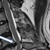 Cervical spondylotic myelopathy (CSM) is a common cause of spinal cord dysfunction. The degenerative disease can cause serious neurological symptoms that can significantly affect the quality of life. The heterogenous presentation of CSM and a lack of a single comprehensive outcome instrument can make the management of suspected CSM very difficult. Despite the advances in surgical techniques to treat CSM, the indications to do surgery remain contr... Cervical spondylotic myelopathy (CSM) is a common cause of spinal cord dysfunction. The degenerative disease can cause serious neurological symptoms that can significantly affect the quality of life. The heterogenous presentation of CSM and a lack of a single comprehensive outcome instrument can make the management of suspected CSM very difficult. Despite the advances in surgical techniques to treat CSM, the indications to do surgery remain contr...
| |
Open Access DOI:10.23937/2643-4474/1710055 Primary Intramedullary Frontal Bone Osteoblastoma: A Case Report Senai Goitom Sereke, MBChB, MMed (Radiology) and Felix Bongomin, MBChB, MSc Article Type: Case Report | First Published: January 16, 2021  Osteoblastoma is a rare osteoid tissue forming tumor commonly affecting the posterior element of the spine. Intramedullary osteoblastoma of the calvarium is extremely rare. Osteoblastoma rarely occurs in the first decade of life. In this report we describe osteoblastoma of a very uncommon location in an 8-years-old male. An 8-year-old male presented with a 2-year history of progressive painless swelling of the left forehead. There was no prior hi... Osteoblastoma is a rare osteoid tissue forming tumor commonly affecting the posterior element of the spine. Intramedullary osteoblastoma of the calvarium is extremely rare. Osteoblastoma rarely occurs in the first decade of life. In this report we describe osteoblastoma of a very uncommon location in an 8-years-old male. An 8-year-old male presented with a 2-year history of progressive painless swelling of the left forehead. There was no prior hi...
| |
Open Access DOI:10.23937/2643-4474/1710054 Ryuzaburo Kanazawa, Takanori Uchida, Tetsuhiro Higashida, Yuichi Takahashi, Naoyuki Arai, Hidenori Ohbuchi and Tomoyuki Yoshihara Article Type: Technical Note | First Published: December 31, 2020 In some endovascular procedures, the insertion of stent delivery catheters (SDCs) is difficult. We demonstrated the efficacy of the Echelon 10 MicroCatheter (90-degree angle; Medtronic, Dublin, Ireland) during such procedures. We used the Echelon 10 MicroCatheter during difficult procedures to navigate SDCs to the distal artery from the aneurysm. The purpose of this was to reduce mechanical stress on the aneurysmal wall. The SDC was inserted by means of an exchange technique with an intermediate...
| |
Open Access DOI:10.23937/2643-4474/1710053 Use of Acetazolamide in the Postoperative Care of Endoscopic Cerebrospinal Fluid Leak Closure J Nuyts, J Demeestere, M Jorissen and L Van Gerven Article Type: Case Report and Literature Review | First Published: December 31, 2020 Spontaneous cerebrospinal fluid (CSF) leaks are defined as leaks without identifiable etiology, often presenting as spontaneous rhinorrhea. This is thought to be a variant of idiopathic intracranial hypertension (IIH). Spontaneous CSF leak closure often requires surgery, which has proven to be less successful than their non-spontaneous counterparts. There is growing evidence that active intracranial pressure (ICP) management in the postoperative care can improve success rates. We aim to give an ...
| |
Open Access DOI:10.23937/2643-4474/1710052 Segmental Spinal Dysgenesis: Case Report and Literature Review Alejandro Vargas-Moreno, María Fajardo-Montes, Diego Miguel Rivera-Mariño and Juan Carlos Acevedo-González Article Type: Case Report and Literature Review | First Published: December 26, 2020 Full-term female-neonate, delivered by cesarean section without complications, with prenatal ultrasound documentation of bilateral congenital talipes equinovarus and suggestive imaginological signs of a non-specific thoracolumbar spine malformation. At the time of birth, the physical examination reflected the presence of a dorsal complex cutaneal appendix covered by skin, associated with lower limbs hypertonia and bilateral talipes equinovarus. There were no respiratory alterations. MRI images o...
| |
Open Access DOI:10.23937/2643-4474/1710051 Spontaneous Resolution of Sub-Acute Subdural Haematoma Krishan Kumar Sharma and Asgar Ali Article Type: Case Report | First Published: December 26, 2020 As per Guidelines for surgical management of Traumatic Brain Injury by Brain Trauma Foundation & The Congress of Neurological surgeons, surgery is indicated in Acute subdural haematoma if CT scan shows midline shift > 5 mm, or haematoma thickness > 10 mm regardless of patient’s Glasgow coma scale score. However, nothing specified separately for sub-acute subdural haematoma. Mostly same criteria are being followed for selecting patients for surgery in sub-acute and chronic subdural haematoma. S...
| |
Open Access DOI:10.23937/2643-4474/1710050 De Novo Cavernoma in the Sensory Cortex Causing Cervicobrachialgia Dhruv Jain, MS, Michael Broduhn, MD, Sophie Rosahl and Steffen K Rosahl, MD, PhD Article Type: Case Study and Review of Literature | First Published: December 17, 2020 A 32-year-old female presented with neck pain radiating to the ulnar forearm along with occasion-al tingling sensations. Three years earlier, she had suffered from left-sided cerebellar infarction caused by dissection of the left vertebral artery. The dissection was provoked by manual therapy administered by a certified physiotherapist. Antiplatelet treatment with Aspirin 100 mg/day was established. One year later, she experienced tingling in the left side of her face. At the time, intracerebral...
| |
Open Access DOI:10.23937/2643-4474/1710049 Orbital Apex Syndrome and Carotid Artery Injury Associated with Transorbital Stab Injury Dimitri Laurent, MD, Brian Corlis, MD and Gregory JA Murad, MD Article Type: Case Report | First Published: December 09, 2020 Transorbital penetrating injuries are particularly rare in the adult population. We present a case of TOPI sustained in an assault using a ballpoint pen-with concomitant transmaxillary injury due to a pencil stab wound-and a review of the literature. Transorbital penetrating injuries (TOPI) are particularly rare in the adult population, accounting for 0.096% of all head injuries. In the setting of TOPI, there should be a high index of suspicion for damage to the intracranial vascular and neural ...
| |
Open Access DOI:10.23937/2643-4474/1710048 Carlos Roberto Massella, MD, Yasmeen Elsawaf, BS, Sabino Luzzi, MD, Paulo Mácio Porto de Melo, MD and Samer K Elbabaa, MD, FAANS, FAAP, FACS Article Type: Case Report and Comprehensive Literature Review | First Published: December 06, 2020 Resection of a cavernous sinus tumor may be performed utilizing three primary approaches: The middle cranial fossa epidural approach, the combined transsylvian- subtemporal approach, and the medial transsphenoidal approach. Herein, we describe an exceedingly rare cavernous sinus chondroma in an 18-year-old male who presented with a history of binocular diplopia and trochlear nerve palsy. Brain CT scan detected a left sided cavernous sinus calcified mass and the MRI confirmed an enhancing lesion ...
| |
Open Access DOI:10.23937/2643-4474/1710047 Crystal N Adams, MD, Mohamad Z Koubeissi, MD, Aalap Herur-Raman and Donald C Shields, MD, PhD Article Type: Case Report | First Published: November 28, 2020 Deep brain stimulation of the anterior thalamic nucleus for treatment of pharmacoresistant epilepsy is a surgical option for patients who are not candidates for resective surgery. One of the challenges of this technique relates to the small size of the anterior thalamic nucleus (approximately 10 mm × 5 mm × 5 mm). Stimulation several millimeters outside the intended target provides suboptimal seizure control. One option for achieving accurate stimulation includes the use of virtual reality sof...
| |
Open Access DOI:10.23937/2643-4474/1710046 Case Report: A Rare Case of Penetrating Trauma of Frontal Sinus with Anterior Table Fracture Himanshu Raval, Mona Bhatt and Nihar Gaur Article Type: Case Report | First Published: November 25, 2020 Head injury is common component of any road traffic accident injury. Injury involving only frontal sinus is uncommon and unique as its management algorithm is changing over time with development of radiological modalities as well as endoscopic intervention. Frontal sinus injuries may range from isolated anterior table fractures causing a simple aesthetic deformity to complex fractures involving the frontal recess, orbits, skull base, and intracranial contents. Only anterior table injury of front...
| |
Open Access DOI:10.23937/2643-4474/1710045 Chordoid Meningioma: Case Report and Review of the Literature Marek Prokopienko, Teresa Wierzba-Bobrowicz, Wiesława Grajkowska, Tomasz Stępień and Michał Sobstyl Article Type: Case report and Review of the Literature | First Published: November 16, 2020 Chordoid meningioma is an uncommon histopathological type of meningioma, frequently associated with Castleman’s syndrome. Histologically, chordoid meningiomas are similar to chordomas. Because of their high proliferative index, they present aggressive biological behavior and high risk of postoperative recurrence. We report a case of choroid meningioma in adult patient without Castleman’s syndrome manifestation. As its chordoid features is related with a rapid recurrence after incomplete remo...
| |
Open Access DOI:10.23937/2643-4474/1710044 A Case of Subarachnoid Hemorrhage from the Posterior Pial Artery of the Medulla Oblongata Takahiro Ogishima, MD, PhD, Takashi Sugawara, MD, PhD, Shoko Hara, MD, Tadashi Nariai, MD, PhD, and Taketoshi Maehara, MD, PhD Article Type: Case report and Case Series | First Published: October 29, 2020 We present a case with localized subarachnoid hemorrhage in cisterna magna. The patient exhibited re-bleeding after admission. Repeated angiography couldn’t find the bleeding cite. Surgical exploration revealed that the responsible artery was a tiny pial artery on the posterior surface of medulla oblongata. Even though the artery was too small to be delineated in angiogram, subarachnoid hemorrhage and re-bleeding did occur. In this report we discuss the cause of the small artery rupture and po...
| |
Open Access DOI:10.23937/2643-4474/1710043 Chondromyxoid Fibroma of Lumbar Vertebrae: Case Report and Literature Review İsmail KAYA, MD Article Type: Case Report and Literature Review | First Published: October 01, 2020 Chondromyxoid fibroma is a benign tumor of long bone metaphysis. In this article, we reported L4 laminar attachment of chondromyxoid fibroma case and made extensive literature revive. A 56-year-old Caucasian female unable to walk without pain at her right leg diagnosed with chondromyxoid fibroma at right l4 lamina. Complete excision of the lesion via routine lumbar disc surgery with 6 months follow up without pain. Her Hypoesthesia cured also muscle weakness totally recovered. Chondromyxoid fibr...
| |
Open Access DOI:10.23937/2643-4474/1710042 Zhanna Semenova, MD, PhD and Andrei Marshintsev Article Type: Case Report | First Published: September 21, 2020 In the absence of autogenous bone due to various reasons, the choice of an implant becomes an important problem of reconstructive surgery. With introduction of computer modelling and development of prototyping technologies the issues of cosmetic reconstructive operations are becoming less relevant. In late cranioplasty implant production and installation is complicated by the presence of significant bone growth along the perimeter of cranial defect. The ability to adjust implant to a bone window...
| |
Open Access DOI:10.23937/2643-4474/1710041 Multiple Spinal Metastases from a Malignant Paraganglioma Chrystal Calderon, MBBS, MRCS, Alberto Perez Villafuerte, Arianne Lalla, MBBS and Sandeep Maharajh, MBBS Article Type: Case Report | First Published: September 16, 2020 A malignant paraganglioma is a rare form of cancer that depicts varying symptomatology on presentation. This case serves to highlight an aggressive form of the disease, with considerable metastasis to the cervical, thoracic, and lumbar spine. This case was reliant on histopathological analysis to confirm the diagnosis. A 34-year-old male of Afro-Caribbean descent presented with a 1-month history of shortness of breath, body pains and lower limb weakness. On investigation for a pulmonary embolus,...
| |
Open Access DOI:10.23937/2643-4474/1710040 Reconstruction of Cranial Defect With Individualized PEEK Implant: Case Report Cristopher Mayer O, Tomás Donoso H, Roberto García B, Claudio Huentequeo M and Francisco Pampin L Article Type: Case Report | First Published: August 12, 2020 Cranioplasty has numerous indications, and its main objective is to support the underlying noble structures, along with restoring shape and aesthetics. The treatment of choice is controversial, and is mainly related to the type of material to be used. There were no complications in the perioperative period, nor were there rejection reactions, infection or exposure of the implant in the postoperative period, with satisfactory aesthetic (improvement of the cranial contour) and functional results d...
| |
Open Access DOI:10.23937/2643-4474/1710039 Velázquez Domínguez Héctor Eduardo, Guerrero Jazo Francisco Javier, Velázquez Santana Héctor, Pozos Ochoa Luis Iván, López Rodríguez Christopher Josué, Salcedo Hernández Missael de Jesús and Fraga González Rodrigo Article Type: Case Report | First Published: July 27, 2020 Pleomorphic xanthoastrocytoma (PXA) is a rare tumor with favorable prognosis, classified as grade II in the World Health Organization (WHO) and accounts for less than 1% of all astrocytic neoplasm. It is commonly found in childhood and young adults. This tumor has been described as part of the spectrum of Long-Term Epilepsy Associated Tumors (LEAT). The most common location is supratentorial, involving predominantly the temporal lobe. Here we present the case of a 6-year-old female with long-ter...
| |
Open Access DOI:10.23937/2643-4474/1710038 Axis Metastasis, Atlantoaxial Dislocation and Cervical Syringomyelia: A Case Report Ismail Latifaci, MD, Selcuk Gozcu, MD and Ahmet Celal Iplikcioglu Article Type: Case Report | First Published: June 19, 2020 Syringomyelia associated with atlantoaxial dislocation is usually described in patients with complex craniovertebral junction anomaly. However, atlantoaxial dislocation without craniovertebral junction bony anomaly very rarely causes syringomyelia. In this report, we describe a case of syringomyelia associated with atlantoaxial dislocation secondary to metastatic tumour of the axis vertebra. Syringomyelia is characterized by the presence of a fluid filled cystic cavity within the spinal cord. Ce...
| |
Open Access DOI:10.23937/2643-4474/1710036 Ossified Cephalhematoma: How to Differentia it from Intradiploic Hematoma: Case Report Selçuk Gözcü and Ahmet Celal İplikçioğlu Article Type: Case Report | First Published: June 18, 2020 Ossified cephalhematoma is rare and usually occur in newborns due to birth injury. Some ossified cephalhematomas can resorb spontaneously while others require surgery because of cosmetic reasons. Simple excision of new bone is usually sufficient. It should be also differentiated from another clinical entity called intradiploic hematoma which requiers surgery. In reported cases, of infant intradiploic hematoma cases clinical and radiologicial findings are similar to those of ossified cephalhemato...
| |
Open Access DOI:10.23937/2643-4474/1710037 Cerebral and Cerebellar Ischaemia in Pituitary Apoplexy: A Case Report Nimetullah Alper Durmus, Iddi Kassım Mkopi and Ali Kurtsoy Article Type: Case Report | First Published: June 19, 2020 Pituitary apoplexy followed by cerebral ischaemia is very rare and life-threatening clinical syndrome. Ischemia is a rare complication after pituitary apoplexy. Cerebral ischemia due to pituitary apoplexy has been linked to two mechanisms; this can occur through one of two mechanisms- direct compression of the artery or vasospasm caused by vasoactive factors/agents released from haemorrhagic or necrotic material of the pituitary adenoma....
| |
Open Access DOI:10.23937/2643-4474/1710034 Management and Outcome of Craniofacial Fibrous Dysplasia in a Tertiary Neurosurgical Centre, Nigeria Nasiru Jinjiri Ismail, Ali Lasseini, Aliyu Muhammad Koko and Bello B Shehu Article Type: Research Article | First Published: May 16, 2020 Fibrous dysplasia is an aberration in normal bone development that results from a defect in osteoblastic differentiation and maturation originating in a mesenchymal precursor; it’s characterized by foci of abnormal fibro-osseous proliferation that can affect any area of the calvaria. Craniofacial fibrous dysplasia is a rare condition that can presents with craniofacial swelling and disfigurement. Usually, cranial computerised tomography scan is needed for radiological evaluation and excision a...
| |
Open Access DOI:10.23937/2643-4474/1710035 Dura Based Rhabdoid Glioblastoma Masquerading as Meningioma: A Rare Case Report Baliga V, Ganapathy S and Pandey P Article Type: Case Report | First Published: May 16, 2020 There are quite a few entities which can present as dura- based masses. Most common being meningioma, others are gliosarcomas, solitary fibrous tumor, leiomyosarcoma, haemangiopericytomas and melanocytomas. Recently, a number of neoplastic and non-neoplastic entities have been reported that radiographically and clinically mimic meningiomas. This is a rare case of glioblastoma with rhabdoid features presented as dura-based tumor. Even though the imaging helps to narrow down the diagnostic possibi...
| |
Open Access DOI:10.23937/2643-4474/1710033 The Imaging Features of AIDS-Related Primary CNS Lymphoma; Case Series and Literature Review Shunsuke Yanagisawa, Yuya Sakakura, Shota Ozaki, Ryuichi Noda, Yuta Tamai, Makiko Fujitani, Masato Inoue and Tetsuo Hara Article Type: Case Series and Literature Review | First Published: May 01, 2020 Primary CNS lymphoma is rare primary brain tumor. Meanwhile there are given number of acquired immune deficiency syndrome related primary CNS lymphoma. This brain tumor was reported that it showed ring enhancement in contrast MRI or CT. Here we analysed six acquired immune deficiency syndrome related primary CNS lymphoma cases. All cases showed ring enhancement in contrast MRI. The central low uptake and peripheral high uptake were observed in 18F-fluorodeoxyglucose PET, and these imaging featur...
| |
Open Access DOI:10.23937/2643-4474/1710032 Management Options in Patients with Trapped Temporal Horn: Case Report and Critical Review Joel Caballero-García, Carlos Aparicio-García, Misael López-Sánchez and Jhohana Linares Benavides Article Type: Case Report | First Published: April 18, 2020 A 58-year-old woman with a previous history of partial resection for a parietal glioblastoma in another center and trapped temporal horn is presented. A ventriculoperitoneal shunt was done with completely resolution of the temporal lobe entrapment. Trapped temporal horn is a rare condition, and many different surgical approaches have been described. Ventriculoperitoneal shunting remains the standard treatment and has some particularly advantages in patients with malignant tumors. The ideal treat...
| |
Open Access DOI:10.23937/2643-4474/1710031 Cranial Intraosseous Angiolipoma: Case Report and Literature Review Katherine M Morgan, MD, PhD, Simon Hanft, MD and Zhenggang Xiong, MD, PhD Article Type: Case Report | First Published: April 08, 2020 Angiolipomas are slow-growing, soft tissue tumors consisting of mature adipocytes and thin-walled blood vessels. While most Angiolipomas are subcutaneous lesions in the trunk and upper extremities, intraosseous angiolipomas are rare at cranial site. We present the case of a 61-year-old female with an enlarging lesion in the left frontoparietal skull following minor head trauma. Radiography confirmed an expansile, enhancing, spiculated bony lesion in the left frontoparietal calvarium with extensi...
| |
Open Access DOI:10.23937/2643-4474/1710030 An Unusual Case of Paraspinal Schwannoma Causing Undiagnosed Low Back Pain Ismail Kaya, MD Article Type: Case Report | First Published: March 24, 2020 Paraspinal schwannomas are relatively common but hardly classified subject of spinal neurosurgery. We present a rare and unclassified case of paravertebral schwannoma that causes non-discogenic low back pain. A 45-year-old Caucasian female came to our clinic with low back pain. Her pain steadily worsened for two years. Her physical exam showed right straight leg raise positivity at 45-degree angle but provocation test negative. Magnetic resonance imaging showed a mass in the paravertebral muscle...
| |
Open Access DOI:10.23937/2643-4474/1710029 AA Sufianov, SM Karasev, RR Khafizov and RA Sufianov Article Type: Case Report | First Published: November 04, 2019 Carotid - cavernous fistulas (CCF) are spontaneous or acquired communications between the internal carotid artery (ICA) and the cavernous sinus, which can be classified as direct or indirect. Direct fistulas between the internal carotid artery and the cavernous sinus can occur due to trauma, ruptures of intracavernous carotid artery aneurysms, collagen deficiency syndromes, stratification of arteries, fibromuscular dysplasia, and direct surgical trauma. Symptoms caused by CCF are associated with...
| |
Open Access DOI:10.23937/2643-4474/1710028 A Review of Radiographic Imaging Findings of Ependymal Tumors Maria Habib Hanna, MD, Bansal A, MD and Belani P, MD Article Type: Review Article | First Published: October 24, 2019 Ependymomas are glial tumors that typically arise from the lining of the ventricles or the central canal of the spinal cord. The most common site of occurrence is within the posterior fossa. Subtypes of ependymomas include anaplastic ependymoma, myxopapillary ependymoma, and subependymoma. Its characteristic imaging features include findings of a heterogeneous mass with necrosis, calcifications, cystic changes, and hemorrhage. Treatment options includes partial resection with or without irradiat...
| |
Open Access DOI:10.23937/2643-4474/1710027 AA Sufianov, SM Karasev, RR Khafizov and RA Sufianov Article Type: Case Report | First Published: October 21, 2019 Arteriovenous abnormalities of cerebral vessels are quite common. One of the main manifestations of this pathology are arteriovenous malformations. Also, a combination of malformations with aneurysmal expansion of the vascular wall of various localization is not rare. The frequent localization of such aneurysms are afferents going to the malformation site; therefore, such aneurysms are also called proximal flow-related aneurysms. As a rule, a combined approach is used mainly for endovascular tre...
| |
Open Access DOI:10.23937/2643-4474/1710026 Cerebellar Liponeurocytoma Presenting with Fatal Tumor Hemorrhage Elia Guadagno, MD, Lorenzo Ugga, MD, Annarita Gencarelli, MD, Valentina Orlando, MD, Sergio Corvino, MD, Maria Rosaria Cervasio, MD, Sara Pignatiello, MD, Andrea Elefante, MD, Francesco Maiuri, MD and Maria Laura Del Basso De Caro, MD Article Type: Case Report | First Published: October 18, 2019 Liponeurocytoma is a very rare tumor of the nervous system, with only 63 reported cases, mainly in the cerebellum and ventricular system. The key-points for the diagnosis are the histological findings of neurocytic cells with lipidic accumulation, with positivity for S-100 and synaptophysin and negativity for IDH-1, and the MR finding of hyperintensity areas on T1Wi corresponding to fatty tissue within the tumor. This article reports a case of cerebellar liponeurocytoma presenting with acute ict...
| |
Open Access DOI:10.23937/2643-4474/1710025 Pharyngeal Perforation after Anterior Cervical Fusion Surgery Munibe Busra Erdem, MD, Emrah Celtikci, MD, Utku Aydil, MD, Mesut Emre Yaman, MD and Hakan Emmez, MD Article Type: Case Report | First Published: October 07, 2019 Pharyngeal perforation is a serious complication of anterior cervical fusion surgery. If it is not diagnosed in the early period and the treatment is delayed, catastrophic consequences may occur which may cause death such as septicemia, mediastinitis, pneumonia and meningitis. We shared the presentation of our two cases. In the first case, pharyngeal perforation and spontaneous extrusion of the C2-C3 fixation device occurred after eight years from surgery. The fixation device removed and the pha...
| |
Open Access DOI:10.23937/2643-4474/1710024 Ibiwari Pepple and Debasish Hajra Article Type: Case Report | First Published: October 05, 2019 Small cell lung cancer (SCLC) accounts for 10-20% of all lung cancers and is characterised by early and rapid spread. Ten percent of patients already have cerebral metastases at time of presentation. We report a case of a 37-yr-old female whose pre-operative CT and MRI brain revealed multicentric left frontal and temporal lesions which mimicked intracerebral abscess intraoperatively and confirmed on gram stain as moderate growth of white cells. The patient was commenced on prolonged antimicrobia...
| |
Open Access DOI:10.23937/2643-4474/1710023 Tension Pneumocephalus Causing Cerebral Venous Sinus Thrombosis Nissar Shaikh, Gulzar Hussain, Adnan Khan, Muhammad Mohsin Khan, Mamdouh Al Mustafa, Moad Ehfeda, Muhammad Zubair and Umais Momin Article Type: Case Report | First Published: September 13, 2019 A 38-year-old Asian male had craniotomy with excision of a left frontal mass which extended into the lateral ventricles. External ventricular and subdural drains were inserted at the end of the procedure. He received mannitol and furosemide intraoperatively. Immediately after surgery he developed tension pneumocephalus, diabetes insipidus and fever. Two days after surgery, magnetic resonance imaging (MRI) showed transverse, sagittal sinus thrombosis extending into the right internal jugular vein...
| |
Open Access DOI:10.23937/2643-4474/1710022 Hu Liang Low and Ahsan Taqvi Article Type: Case Report | First Published: September 05, 2019 Post-traumatic conus medullaris syndromes (CMS) such as after L1-burst fractures are uncommon, being encountered in only 1.7% of patients with spinal cord injuries. These injuries are almost invariably associated with bladder, erectile or bowel dysfunction, disturbance of perineal sensation and varying sensorimotor deficits in the legs. To date, there are only 6 reported cases of CMS after a L1-burst fracture without sensory or motor deficits in the lower limbs. In this case report, we describe ...
| |
Open Access DOI:10.23937/2643-4474/1710021 Andrea Bianco, Riccardo Fornaro, Christian Cossandi, Sara Forgnone, Emanuela Crobeddu and Piergiorgio Car and Gabriele Panzarasa Article Type: Commentary | First Published: July 05, 2019 Pregnancy can change the behavior of brain gliomas with increased growth rate, increased frequency of seizures, and anaplastic transformation. Very rarely the presentation may be hyperacute due to intratumoral hemorrhage. We report and discuss them a management of the hemorrhagic infarction of a newly diagnosed postcentral anaplastic astrocytoma in a pregnant woman. A first MRI discovered the glioma; two days later, after acute neurological deterioration, a second MRI showed a dramatic increase ...
| |
Open Access DOI:10.23937/2643-4474/1710020 Caring for Meningiomas: A Multi-Disciplinary Approach Uri P Hadelsberg, MD and Nevo Margalit, MD Article Type: Commentary | First Published: June 29, 2019 This patient was admitted to our department and eventually underwent surgery with gross total resection after pre-operative endovascular embolization of the tumor. The patient underwent a successful recovery and was discharged without major neurological deficits to rehab. On follow up the patient is doing well and is back home with her children. On the day of surgery the patient was intubated and underwent cerebral embolization with occlusion of several tumor-feeding arteries. The patient was th...
| |
Open Access DOI:10.23937/2643-4474/1710019 Multidirectional Spontaneous Migration of Intracranial Bullet: A Case Report and Literature Review Matías Negrotto, MD, Ramon Figueroa, MD and Katherine Sotomayor, MD Article Type: Case Report | First Published: June 26, 2019 Although gunshot injury to the head is usually mortal, survivors frequently show retained metal shrapnel or full bullet intracranially. An important though uncommon complication of retained bullet is that of spontaneous migration. A migrating intracranial bullet is a dilemma to neurosurgeons, as its management is challenging, since the removal of a deep-seated bullet may cause additional neurological deficit. Migration of a retained bullet may cause damage to vital structures and cranial nerves,...
| |
Open Access DOI:10.23937/2643-4474/1710018 Clinical Performance of a Novel Fully Synthetic Dura Substitute Umang Khandpur, BS, Wilson Z Ray, MD and Matthew R MacEwan, PhD Article Type: Case Series | First Published: June 17, 2019 A retrospective case series was performed to evaluate the efficacy of a novel, fully synthetic dura substitute. Clinical outcomes (e.g. infectious, reoperation rates, cerebrospinal fluid leak) and surgeon experiences with the dura substitute intraoperatively were collected. All methods were approved by the Institutional Review Board at Washington University School of Medicine....
| |
Open Access DOI:10.23937/2643-4474/1710017 The Role of Neurosurgery Mission Trips in Third World Countries Emil Zhalmukhamedov Article Type: Review Article | First Published: June 17, 2019 A summary of available data on neurosurgical mission trips has been collected from PubMed and Google Scholar on the following mesh keywords: Neurosurgery trips, mission neurosurgery, and neurosurgery in third world countries. We gathered our information that was closely related to neurosurgical education such as proficiency, equipment, ethics and outcomes....
| |
Open Access DOI:10.23937/2643-4474/1710016 Psychogenic Hemiplegia after Cervical Spine Discectomies and Fusion Siavash S Haghighi Article Type: Case Report | First Published: June 08, 2019 A case report of psychogenic hemiplegia following a two level anterior cervical discectomies and fusion was contradicted by a thorough electrophysiological findings. To report an unusual case of post-operative hemiplegia in spite of normal electrophysiological and noncompromising radiographic findings. Conversion disorder with sensory or motor system symptoms is a subtype which includes symptoms such as impaired motor balance, paraplegia, muscle weakness, and urinary retention....
| |
Open Access DOI:10.23937/2643-4474/1710015 Delayed Quadriplegia after Cervical Discectomy and Fusion Surgery Siavash S Haghighi, MD, Bertha Ramirez, MD and Richard Zhang, MD Article Type: Case Report | First Published: May 08, 2019 Acute or delayed paraplegia or quadriplegia following anterior cervical discectomies and fusion are not common. We report this single case report of delayed quadriplegia following an anterior cervical discectomies and fusion without any change of sensory or motor evoked potentials during the operation. The intraoperative somatosensory (SSEPs) were performed by stimulation of tibial nerves or the median (MN). Motor evoked potentials (TcMEPs) were recorded from intrinsic hand or foot muscles after...
| |
Open Access DOI:10.23937/2643-4474/1710014 Thierry Patrice, MD, PhD, Romain Bourcier, MD, Hubert Desal, MD, PhD, Bertrand Rozec, MD, PhD and Yvonnick Blanloeil, MD Article Type: Case Study | First Published: April 26, 2019 Oxidative stress (OS) could be involved during intracranial aneurysms (ICA) progression but knowledge about comorbidities likely to induce OS is poor. We studied the medical and surgical history of patients with an ICA discovered after a subarachnoidal haemorrhage (SAH) or unruptured (UIA). 175 patients, 58 with a UIA, had been successively recruited from a single centre. Their medical history, comorbidities and treatments had been gathered from hospital files, general practitioners, relatives a...
| |
Open Access DOI:10.23937/NCR-2017/1710013 Self Manipulated Cervical Spine Leads to Posterior Disc Herniation and Spinal Stenosis Wyatt McGilvery, BS, Marc Eastin, MD, Anish Sen, MD and Maciej Witkos, MS, MD Article Type: Case Report | First Published: April 05, 2019 The authors report a case in which a 38-year-old male who presented himself to the emergency department with a chief complaint of cervical neck pain and paresthesia radiating from the right pectoral region down his distal right arm following self manipulation of the patient's own cervical vertebrae. Initial emergency department imaging via cervical X-ray and magnetic resonance imaging (MRI) without contrast revealed no cervical fractures; however, there was evidence of an acute cervical disc her...
| |
Open Access DOI:10.23937/NCR-2017/1710012 Cerebello-Pontine Angle Glioblastoma with Cervical Spine Metastasis: A Case Report Giuseppe Mariniello, MD, Carmela Peca, MD, Maria Laura Del Basso De Caro, MD, Sergio Corvino, MD, Valentina Orlando, MD, Elia Guadagno, MD and Francesco Maiuri, MD Article Type: Case Report | First Published: February 14, 2019 This article reports a left cerebellopontine angle glioblastoma presenting with two week history of rapidly progressive hearing loss and trigeminal pain, treated by subtotal resection and radiotherapy with concomitant and adjuvant temozolomide. At one year, local tumor control and diffuse neoplastic seeding in the cervical spinal cord were evidenced. Glioblastomas involving the cerebellopontine angle are exceptional with only 10 reported cases. The possibility of a glioblastoma should be conside...
| |
Open Access DOI:10.23937/ncr-2017/1710011 Pouya Entezami, MD, Emad Nourollahzadeh, MD, Adedamola Adepoju, MD, David Michael Jones, MD and John Dalfino, MD Article Type: Case Report | First Published: February 11, 2019 As our patient population grows older with improved health care technology and resources, chronic subdural hematoma (CSDH) is an increasingly common disease faced by the modern neurosurgeon. Despite good results with burr-hole irrigation and evacuation - the gold standard treatment for CSDH - recurrence rate remains high. Recent advancements in endovascular management of this diagnosis via embolization of the middle meningeal artery (MMA), either alone or in conjunction with surgical evacuation,...
| |
Open Access DOI:10.23937/ncr-2017/1710010 Delayed Revascularization of Basilar Artery Occlusion due to Symptomatic Progression Pouya Entezami, M Reid Gooch, Emad Nourollahzadeh and John Dalfino Article Type: Case Report | First Published: January 21, 2019 The basilar artery is the main arterial contributor to the posterior circulation. Interruptions of this flow can cause devastating strokes and neurological demise. While most patients with a basilar artery occlusion suffer from a high rate or morbidity and/or mortality, a small subset survive but have progressive symptomatic decline. Delayed revascularization of these patients with chronic basilar artery occlusions based on symptomatic progression may provide a favorable risk-benefit ratio, but ...
| |
Open Access DOI:10.23937/ncr-2017/1710009 Alexandra Benouaich-Amiel, Shlomit Yust-Katz, Suzana Fichman and Tali Siegal Article Type: Case Report | First Published: December 20, 2018 Lymphomatoid granulomatosis (LYG) is a rare, Epstein barr virus (EBV) associated, angiocentric B cell proliferation, which usually involves the lung but may also involve, more rarely, the central nervous system. Its malignant potential is uncertain, and seems to be linked to the number of EBV positive B cells. We report the case of a 32-years-old patient, with an antecedent of LYG grade 2, involving lung, who presented with a brain mass lesion one year later, which appears to be a grade 1 LYG wi...
| |
Open Access DOI:10.23937/ncr-2017/1710008 Multimodal MRI of Extracranial Glioblastoma Dissemination Alice Venier, Luca Roccatagliata, Alessandro Cianfoni and Emanuele Pravata Article Type: Case Report | First Published: December 15, 2018 Glioblastoma scalp dissemination is uncommon. Infiltration may occur through the craniotomy, suggesting diffusion from the surgical site as the most likely mechanism. At Magnetic Resonance Imaging (MRI), features of the metastatic tissue resemble those of the primary tumor. We show multimodal MRI appearance of a glioblastoma disseminating to the scalp. The patient presented with a stiff, non-tender palpable mass within the extracranial soft tissues, five months after surgery. Distinctive finding...
| |
Open Access DOI:10.23937/ncr-2017/1710007 A Giorgianni, P Veiceschi, G Agresta, S Balbi and D Locatelli Article Type: Case Report | First Published: November 19, 2018 We report a case of right internal carotid artery pseudo-aneurysm, arising in the aspects of a giant macroadenoma in a 57-years-old man, presented with right severe temporal orbital headache and vomiting. Computed tomography scan (CT) revealed a destructive skull base mass extended from sellar region to clivus. Pre and post-gadolinium brain magnetic resonance (MRI) confirmed the lesion, characterized by an encasement of optic nerves and carotid siphons bilaterally, with a pseudo-aneurysm of cave...
| |
Open Access DOI:10.23937/ncr-2017/1710006 Adriana Rodrigues Liborio dos Santos, Gabriel Mufarrej, Priscila Oliveira da Conceicao, Paulo Luiz da Costa Cruz, Daniel Dutra Cavalcanti, Leila Chimelli and Paulo Niemeyer Filho Article Type: Original Article | First Published: November 19, 2018 Selective Amygdalohippocampectomy (SAH) is a widespread technique for Mesial Temporal Lobe Epilepsy (MTLE) treatment. Dr. Niemeyer was the first to describe SAH using transventricular approach technique in 1958. In 2018, we celebrate 60 years of the original description of Niemeyer's approach. This study reviews the approach in light of currently technology and shows the results achieved with patients submitted to SAH following Niemeyer's approach at Instituto Estadual do Cérebro Paulo Niemeyer...
| |
Open Access DOI:10.23937/ncr-2017/1710005 Frazier Suction Cannula Fluid Control Technical Note Manuel Dujovny, Channan Kositzke, Pablo Sosa and Fabian Cremaschi Article Type: Research Article | First Published: November 10, 2018 Suction capability in neurosurgery is a critical tool utilized to clear the field and improve visualization of anatomical structures. It is also used for fragmentation and debulking of pathological tumor masses. Prior to the Frazier cannula, there was no orifice bent pressure relief valve associated with suction; this tool provides independent dual suction force. The Frazier cannula has the capability to reach absolute suction or zero suction instantaneously. In this article, we introduce a new ...
| |
Open Access DOI:10.23937/ncr-2017/1710004 Vaner Koksal Article Type: Case Report | First Published: November 05, 2018 Acute peritentorial subdural hematoma (APSDH) is an extremely rare intracranial bleeding, which generally occurs due to a trauma. And, It usually regresses spontaneously without the need for a surgical intervention. However, it is difficult to determine the surgical method when it is life-threatening. A 15-year-old boy was brought to the hospital after a crash with a car whilst on a motorcycle. No significant intracranial bleeding was detected following an isolated severe head trauma; however, G...
| |
Open Access DOI:10.23937/ncr-2017/1710003 What Should Guide Decision-Making Process in Urgency? A Curious Case of Right Acute Hemiparesis Francesco Restelli, Gianluca Agresta, Tommaso Alfiero, Alessandro Dario and Davide Locatelli Article Type: Case Report | First Published: October 31, 2018 As physicians and neurosurgeons, our daily practice in Emergency Departments sometimes calls us to manage patients and to take decisions in sub-optimal conditions, maybe with partial anamnesis, incomplete neurological examinations or with lacks in routine radiological and laboratory exams. Such eventuality, although it may sometimes lead to misdiagnose and to confound some pathological conditions, fortunately is not always linked to bad repercussions for patients. This is especially true when de...
| |
Open Access DOI:10.23937/ncr-2017/1710002 A Giant Cell Granuloma of the Temporal Bone: A Case Report Giyas Ayberk, Selen Isık Adiloglu, Aylin Kılıc Yazgan, Mahmut Ferhat and Mehmet Faik Ozveren Article Type: Case Report | First Published: June 15, 2018 A giant cell granuloma (GCG) rarely occurs in the skull bones. Although it has destructive character, the GCG is used as the synonym of giant cell reparative granuloma (GCRG) in the literature. Differentiation from other osteolytic lesions quite difficult. The first case of GCG in the temporal bone was reported by Hirschl and Katz in 1974. Even though the GCG is not a true neoplasm, the locally aggressive behavior of this tumor necessitates surgical excision whenever possible....
| |
Open Access DOI:10.23937/ncr-2017/1710001 Primary Central Nervous System Lymphoma: A Differential of Midline Crossing Lesions Renu Pokharna, Miles W Reese, Souvik Sen and Tushar Trivedi Article Type: Case Report | First Published: January 11, 2018 Primary Central Nervous System Lymphoma (PCNSL) is a rare non-Hodgkin type neoplasm, which crosses the midline. We report an unusual case of a 71-year-old Caucasian female who was shown to have PCNSL by a tissue biopsy after the brain Magnetic Resonance Imaging (MRI) showed Central Nervous System (CNS) lesions crossing the corpus callosum....
|
Editor-in-chief

ClinMed Archive
7
5
8
5
Articles Published
All articles are fully peer reviewed, free to access and can be downloaded from our ClinMed archive.
Contact our editorial office

ClinMed Journals Index Copernicus Values
Clinical Medical Image Library: 93.51
International Journal of Critical Care and Emergency Medicine: 92.83
International Journal of Sports and Exercise Medicine: 91.84
International Journal of Womens Health and Wellness: 91.79
Journal of Musculoskeletal Disorders and Treatment: 91.73
Journal of Geriatric Medicine and Gerontology: 91.55
Journal of Infectious Diseases and Epidemiology: 91.55
Clinical Medical Reviews and Case Reports: 91.40
International Archives of Nursing and Health Care: 90.87
International Journal of Ophthalmology and Clinical Research: 90.80
International Archives of Urology and Complications: 90.73
Journal of Clinical Nephrology and Renal Care: 90.33
Journal of Family Medicine and Disease Prevention: 89.99
Journal of Clinical Gastroenterology and Treatment: 89.54
Journal of Dermatology Research and Therapy: 89.34
International Journal of Clinical Cardiology: 89.24
International Journal of Radiology and Imaging Technology: 88.88
Obstetrics and Gynaecology Cases - Reviews: 88.42
International Journal of Blood Research and Disorders: 88.22
International Journal of Diabetes and Clinical Research: 87.97
New Issues